. 2020 Nov 12;65(22):225013.
doi: 10.1088/1361-6560/abb0c5.

Detective quantum efficiency of intensified CMOS cameras for Cherenkov imaging in radiotherapy

Affiliations

Detective quantum efficiency of intensified CMOS cameras for Cherenkov imaging in radiotherapy

Daniel A Alexander et al. Phys Med Biol. .

Abstract

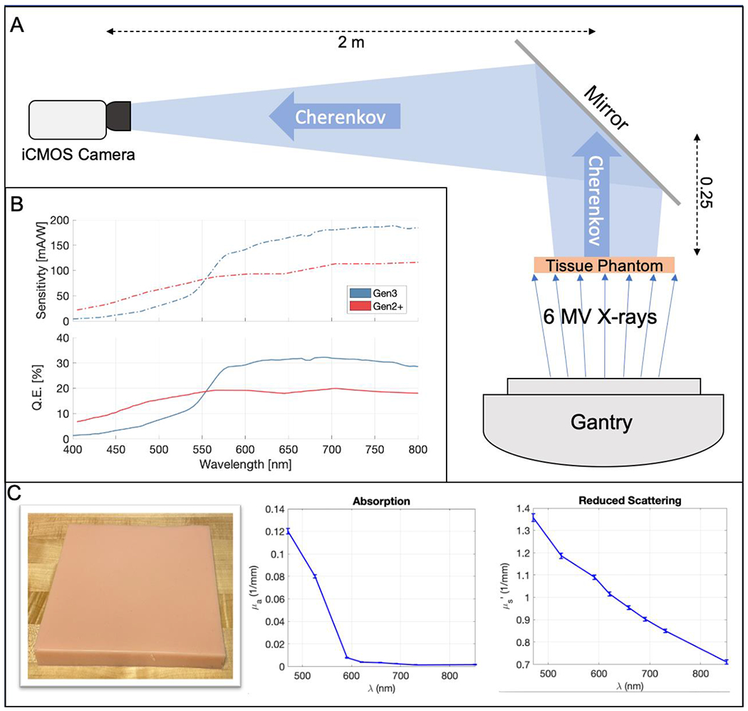
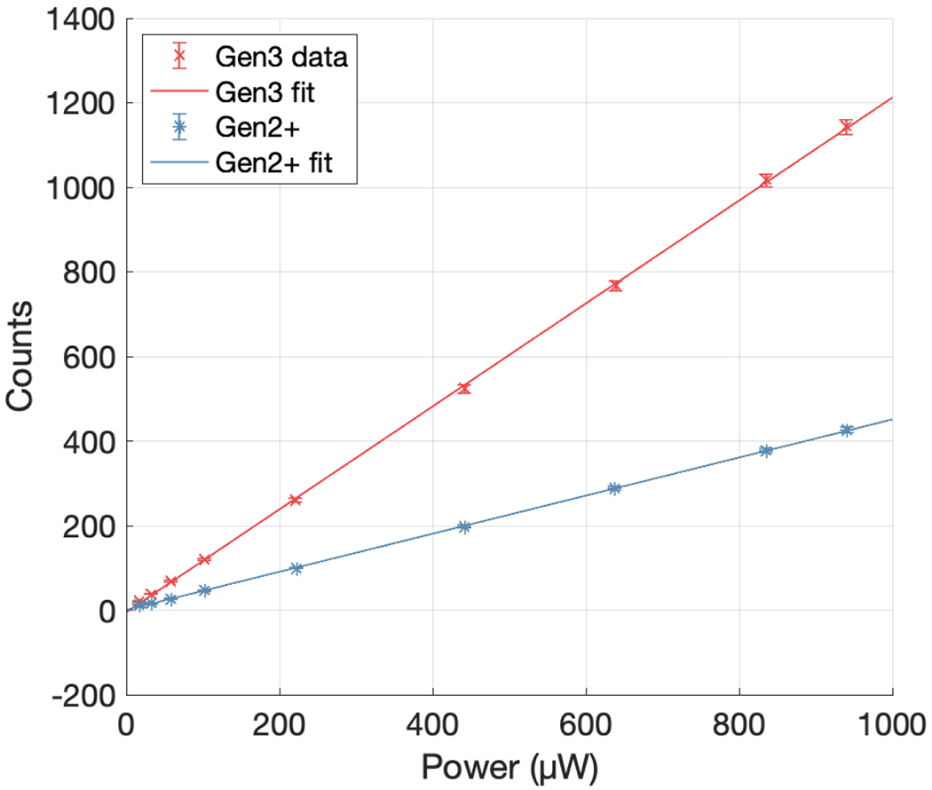
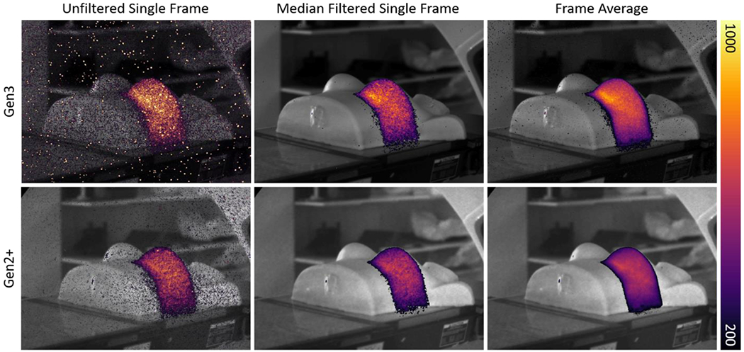
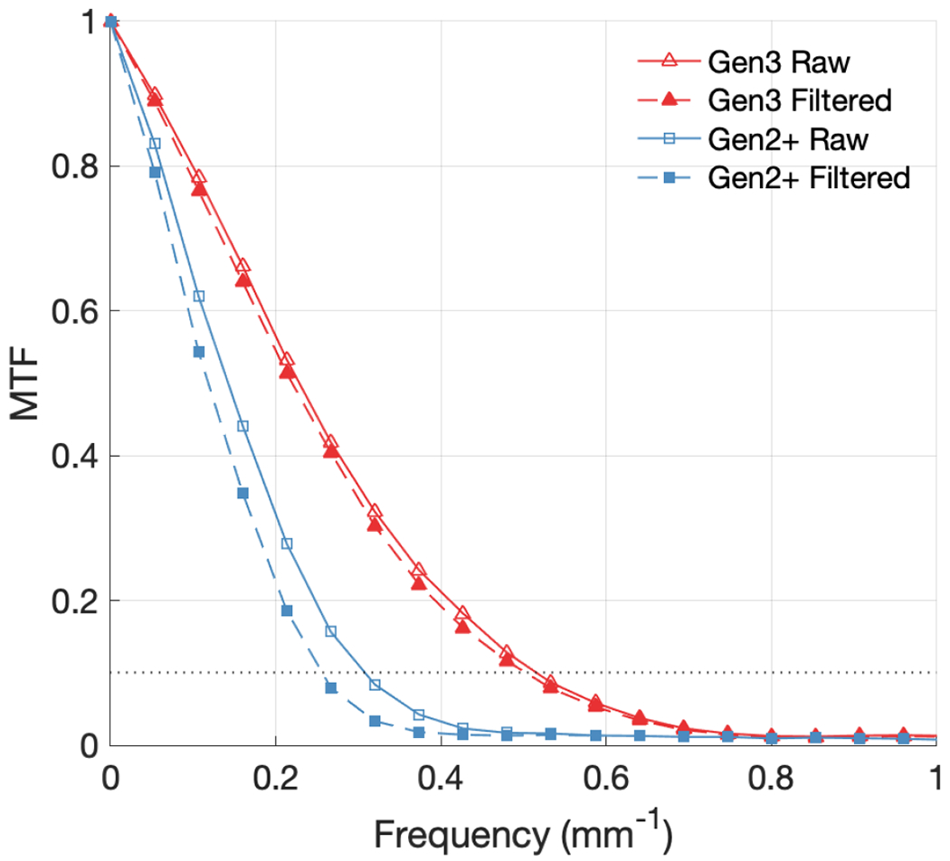
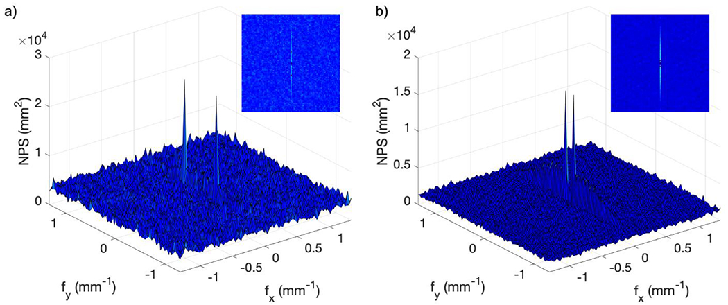
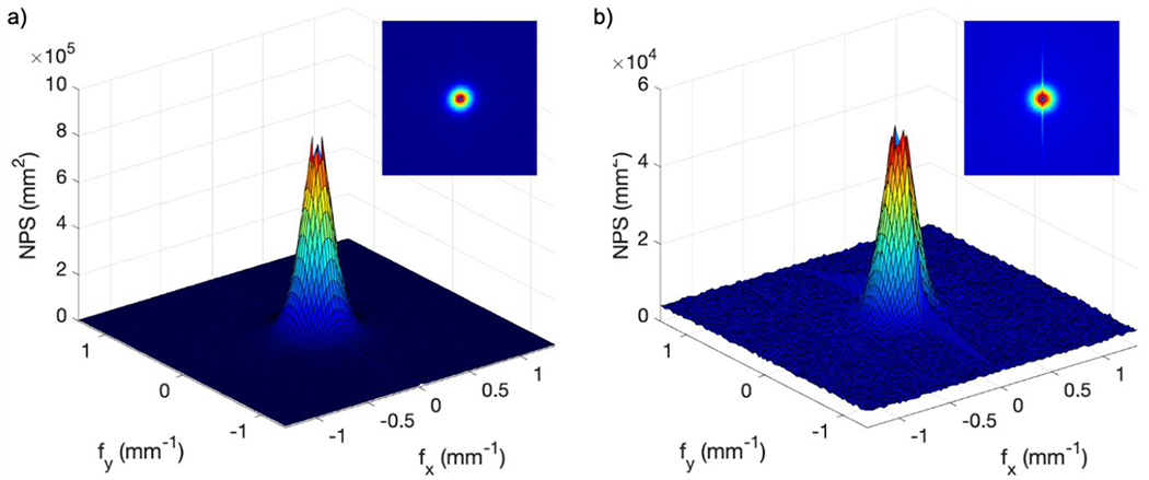
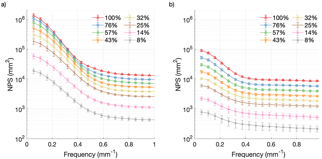
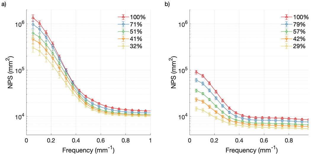
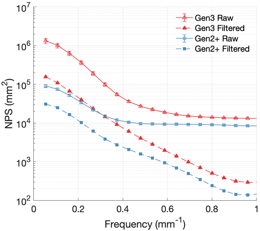
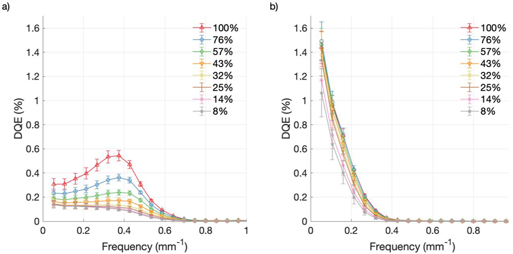
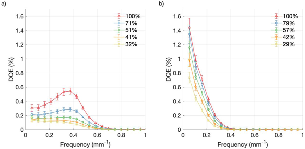
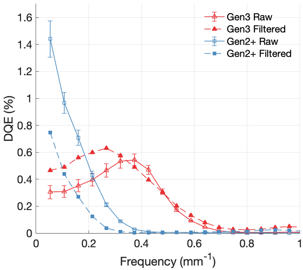
In this study the metric of detective quantum efficiency (DQE) was applied to Cherenkov imaging systems for the first time, and results were compared for different detector hardware, gain levels and with imaging processing for noise suppression. Intensified complementary metal oxide semiconductor cameras using different image intensifier designs (Gen3 and Gen2+) were used to image Cherenkov emission from a tissue phantom in order to measure the modulation transfer function (MTF) and noise power spectrum (NPS) of the systems. These parameters were used to calculate the DQE for varying acquisition settings and image processing steps. MTF curves indicated that the Gen3 system had superior contrast transfer and spatial resolution than the Gen2+ system, with [Formula: see text] values of 0.52 mm-1 and 0.31 mm-1, respectively. With median filtering for noise suppression, these values decreased to 0.50 mm-1 and 0.26 mm-1. The maximum NPS values for the Gen3 and Gen2+ systems at high gain were 1.3 × 106 mm2 and 9.1 × 104 mm2 respectively, representing a 14x decrease in noise power for the Gen2+ system. Both systems exhibited increased NPS intensity with increasing gain, while median filtering lowered the NPS. The DQE of each system increased with increasing gain, and at the maximum gain levels the Gen3 system had a low-frequency DQE of 0.31%, while the Gen2+ system had a value of 1.44%. However, at a higher frequency of 0.4 mm-1, these values became 0.54% and 0.03%. Filtering improved DQE for the Gen3 system and reduced DQE for the Gen2+ system and had a mix of detrimental and beneficial qualitative effects by decreasing the spatial resolution and sharpness but also substantially lowering noise. This methodology for DQE measurement allowed for quantitative comparison between Cherenkov imaging cameras and improvements to their sensitivity, and yielded the first formal assessment of Cherenkov image formation efficiency.


Conflict of interest statement

J. C. Farwell and V. Krishnaswamy are employees and B. Pogue is the president and co-founder of DoseOptics LLC, manufacturing the C-Dose cameras provided for this research. P. Bruza is the principal investigator in SBIR subaward B02463 (prime award NCI R44CA199681, DoseOptics LLC). D. Alexander reports receiving consulting fees from DoseOptics LLC outside of this work.

Figures

Figure 1.
Figure 1.
(A) Schematic of imaging geometry. (B) Spectral sensitivity and quantum efficiency curves for both the Sb-K-Na-Cs Gen2+ and GaAs-O-Cs Gen3 intensifiers. (C) Color image of the tissue phantom, as well as absorption and reduced scattering coefficients as a function of wavelength.
Figure 2.
Figure 2.
Pixel response of both the Gen3 and Gen2+ cameras to variation in incident light with power measured in the emission plane.
Figure 3.
Figure 3.
Cherenkov images of a chest phantom taken with both Gen2+ and Gen3 systems with three different levels of processing showing an unfiltered single frame, spatial and temporal median filtered single frame image, and an average image from all the 100 frames.
Figure 4.
Figure 4.
MTF curves compared for Gen3 and Gen2+ systems, with and without median filtering. The dotted line represents 10% of the MTF.
Figure 5.
Figure 5.
Two dimensional darkfield NPS from raw data: (a) Gen3 system; (b) Gen2+ system.
Figure 6.
Figure 6.
Two dimensional flat illumination NPS from raw data: (a) Gen3 system; (b) Gen2+ system.
Figure 7.
Figure 7.
NPS from raw data at various CMOS gain settings: (a) Gen3 system; (b) Gen2+ system.
Figure 8.
Figure 8.
NPS from raw data at various intensifier gain settings: (a) Gen3 system; (b) Gen2+ system.
Figure 9.
Figure 9.
NPS curves compared for Gen3 and Gen2+ systems, with and without median filtering, for the maximum intensifier and CMOS gain settings used.
Figure 10.
Figure 10.
DQE from raw data at various CMOS gain settings: (a) Gen3 system; (b) Gen2+ system.
Figure 11.
Figure 11.
DQE from raw data at various intensifier gain settings: (a) Gen3 system; (b) Gen2+ system.
Figure 12.
Figure 12.
DQE curves compared for Gen3 and Gen2+ systems, with and without median filtering.

Similar articles

Cited by

References

    1. Jarvis LA, Zhang R, Gladstone DJ, et al. Cherenkov video imaging allows for the first visualization of radiation therapy in real time. Int J Radiat Oncol Biol Phys. 2014;89(3):615–622. doi:10.1016/j.ijrobp.2014.01.046 - DOI - PubMed
    1. Hachadorian R, Bruza P, Jermyn M, et al. Correcting Cherenkov light attenuation in tissue using spatial frequency domain imaging for quantitative surface dosimetry during whole breast radiation therapy. J Biomed Opt. 2018;24(07):1. doi:10.1117/1.jbo.24.7.071609 - DOI - PMC - PubMed
    1. Alexander DA, Tendler II, Bruza P, et al. Assessment of imaging Cherenkov and scintillation signals in head and neck radiotherapy. Phys Med Biol. Published online 2019. doi:10.1088/1361-6560/ab25a3 - DOI - PMC - PubMed
    1. Beddar S, Archambault L, Sahoo N, et al. Exploration of the potential of liquid scintillators for real-time 3D dosimetry of intensity modulated proton beams. Med Phys. 2009;36(5):1736–1743. doi:10.1118/1.3117583 - DOI - PMC - PubMed
    1. Pönisch F, Archambault L, Briere TM, et al. Liquid scintillator for 2D dosimetry for high-energy photon beams. Med Phys. 2009;36(5):1478–1485. doi:10.1118/1.3106390 - DOI - PMC - PubMed

Publication types