Review
. 2020 Sep;112(3):138-152.
doi: 10.32074/1591-951X-164.

Neoplastic and pre-neoplastic lesions of the oesophagus and gastro-oesophageal junction

Affiliations
Review

Neoplastic and pre-neoplastic lesions of the oesophagus and gastro-oesophageal junction

Federica Grillo et al. Pathologica. 2020 Sep.

Abstract

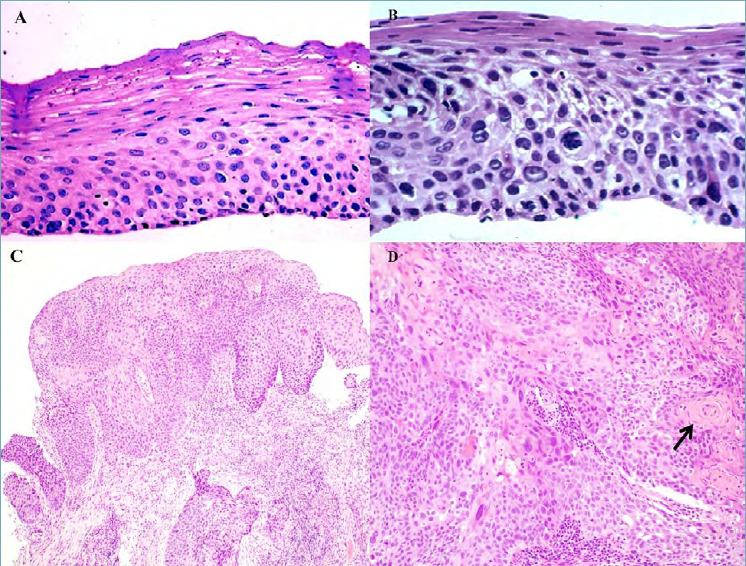
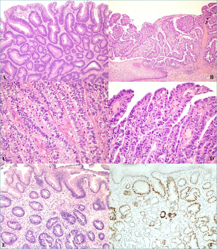
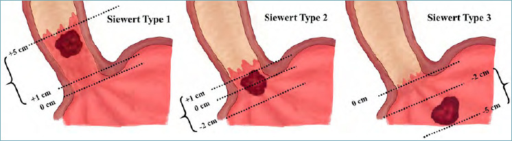
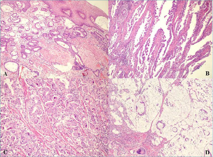
Oesophageal and gastro-oesophageal junction (GOJ) neoplasms, and their predisposing conditions, may be encountered by the practicing pathologist both as biopsy samples and as surgical specimens in daily practice. Changes in incidence of oesophageal squamous cell carcinomas (such as a decrease in western countries) and in oesophageal and GOJ adenocarcinomas (such as a sharp increase in western countries) are being reported globally. New modes of treatment have changed our histologic reports as specific aspects must be detailed such as in post endoscopic resections or with regards to post neo-adjuvant therapy tumour regression grades. The main aim of this overview is therefore to provide an up-to-date, easily available and clear diagnostic approach to neoplastic and pre-neoplastic conditions of the oesophagus and GOJ, based on the most recent available guidelines and literature.

Keywords: Barrett’s dysplasia; oesophageal adenocarcinoma; oesophageal dysplasia; oesophageal squamous cell carcinoma; tumour regression grade.


Conflict of interest statement

Conflict of interest

The Authors declare no conflict of interest.

Figures

Figure 1.
Figure 1.
(A) Oesophageal biopsy section showing low grade squamous dysplasia with only mild cytological atypia within the lower half of the epithelial thickness (magnification x40). (B) Oesophageal biopsy section showing high grade squamous dysplasia with severe cytological atypia in more than half of the epithelium (magnification x40). (C) Dysplastic squamous epitelium and invasive squamous cell carcinoma (magnification x10). (D) Invasive squamous cell carcinoma with keratin pearl formation (arrow) (magnification x20).
Figure 2.
Figure 2.
(A) Intestinal type low grade dysplasia in Barrett’s oesophagus showing scanty architectural distortion and mild to moderate cytological atypia; hyperchromatic nuclei with irregular contours, nuclear overlapping and crowding are seen (magnification x20). (B) Intestinal type high grade dysplasia in Barrett’s oesophagus showing both architectural abnormality and severe cytological atypia adjacent to squamous epithelium (magnification x10). (C) Foveolar type low grade dysplasia in Barrett’s oesophagus with closely packed glands with a single layer of columnar cells with no interspersed goblet cells, round/oval basal nuclei with little stratification or pleomorphism and vesicular nuclei (magnification x20). (D) Foveolar type high grade dysplasia with enlarged cells with greater pleomorphism, loss of polarity and increased mitoses (magnification x20). (E) Crypt dysplasia: dysplasia is confined to the crypt with surface epithelium maturation (magnification x20). (F) p53 immunostained section of crypt dysplasia showing p53 nuclear accumulation in the crypt areas of dysplastic epithelium but not in the surface epitheium.
Figure 3.
Figure 3.
Schematic representation of the Siewert macroscopic classification of gastro-oesophageal junction tumours. Siewert Type I: Adenocarcinoma of the distal oesophagus. The tumour centre is located 1-5 cm above the gastric cardia. Siewert Type II: Adenocarcinoma of the GOJ/cardia. The tumour centre is located 1 cm above or 2 cm below the gastric cardia. Siewert Type III: Adenocarcinoma of the subcardial stomach. The tumour centre is located 2-5 cm below the gastric cardia. In the figure, 0 cm represents the gastric cardia.
Figure 4.
Figure 4.
(A) Invasive oesophageal adenocarcinoma, tubular/glandular type which invades and undermines squamous epithelium (magnification x10). (B) Invasive oesophageal/GOJ adenocarcinoma, papillary type (magnification x10). (C) Invasive oesophageal/GOJ adenocarcinoma with micropapillary features (magnification x20). (D) Invasive oesophageal/GOJ adenocarcinoma, mucinous type (magnification x10).
Figure 5.
Figure 5.
(A) Invasive oesophageal/GOJ adenocarcinoma, poorly cohesive non signet ring cell type (magnification x40). (B) Invasive oesophageal adenocarcinoma, poorly cohesive signet ring cell type (magnification x40). (C) Invasive oesophageal/GOJ adenocarcinoma, undifferentiated carcinoma with lymphoepithelioma-like carcinoma features including a syncytial pattern and prominent lymphocyte infiltration (magnification x40). (D) Invasive oesophageal/GOJ adenocarcinoma, adenosquamous type with squamous areas (asterisks) and glandular areas with mucin production (arrows) (magnification x20).

References

    1. WHO Classification of Tumours of the Digestive System. 5th ed. Geneva: World Health Organization Classification of Tumours. WHO Classification of Tumours Editorial Board. Lyon: IARC press; 2019.
    1. RARECARENet Information Network on Rare Cancers. Accessed: 15 Sept 2019. Available from http://rarecarenet.eu/index.php
    1. Pan Q-J, Roth MJ, Guo H-Q, et al. . Cytologic detection of esophageal squamous cell carcinoma and its precursor lesions using balloon samplers and liquid-based cytologyin asymptomatic adults in Llinxian, China. Acta Cytol 2008;52:14-23. https://doi.org/10.1159/000325430 10.1159/000325430 - DOI - PubMed
    1. Ismail-Beigi F, Horton PF, Pope CE. Histological consequences of gastroesophageal reflux in man. Gastroenterol 1970;58:163-74. - PubMed
    1. Weinstein WM, Bogoch ER, Bowes KL. The normal human esophageal mucosa : a histological rappraisal. Gastroenterol 1975;68:40-4. - PubMed

MeSH terms

Supplementary concepts