Review
. 2020 Sep;112(3):153-165.
doi: 10.32074/1591-951X-163.

Gastritis: update on etiological features and histological practical approach

Affiliations
Review

Gastritis: update on etiological features and histological practical approach

Gianmaria Pennelli et al. Pathologica. 2020 Sep.

Abstract

Gastric biopsies represent one of the most frequent specimens that the pathologist faces in routine activity. In the last decade or so, the landscape of gastric pathology has been changing with a significant and constant decline of H. pylori-related pathologies in Western countries coupled with the expansion of iatrogenic lesions due to the use of next-generation drugs in the oncological setting. This overview will focus on the description of the elementary lesions observed in gastric biopsies and on the most recent published recommendations, guidelines and expert opinions.

Keywords: H. pylori; endoscopy; gastritis; secondary prevention; staging.


Conflict of interest statement

Conflict of interest

The Authors declare no conflict of interest.

Figures

Figure 1
Figure 1
(A-B). Helicobacter pylori active gastritis (Giemsa staining). Lymphocytic inflammation and neutrophilic epithelium infiltration with conventional spiral-shaped H. pylori (A; 630x magnification). Dormant or stressed coccoid microorganisms form (arrows, B; 630 x magnification).
Figure 2.
Figure 2.
(A-B). Autoimmune gastritis. Early phase of autoimmune gastritis with hypertrophic glandular changes and mild lymphocytic and granulocytic infiltrate in the lamina propria (A; 200x magnification). The end stage is characterized by a marked replacement of oxyntic glands with pseudopyloric and intestinal metaplasia with mild inflammation of the lamina propria (B; 100x magnification).
Figure 3.
Figure 3.
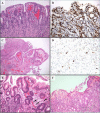
(A-F). Special-type gastrites. Lymphocytic gastritis is defined by the presence of at least 25 intraepithelial lymphocytes CD3+ per 100 gastric epithelial cells (A; hematoxylin-eosin, 200x magnification) (B; CD 3 immunostaining, 200x magnification). Cytomegalovirus (CMV) gastritis is characterized abundant granulation tissue with important inflammatory reaction (C; hematoxylin-eosin, 200x magnification) and CMV inclusions are visible in endothelial cells and also in macrophages (D; CMV immunostaining, 630x magnification). Graft versus host disease (GVHD) gastric mucosa shows apoptotic bodies (arrows) and gland abscess (E; hematoxylin-eosin, 200x magnification). Collagenous gastritis is typically defined by the subepithelial deposition of collagen bands thicker than 10 μm and the intense inflammation response in the lamina propria (F; hematoxylin-eosin, 200x magnification).
Figure 4
Figure 4
(A-B). Enterochromaffin-like cells disorders in autoimmune gastritis. Linear hyperplasia of endocrine cells growing within the gastric gland (double arrow) and micronodular hyperplasia in the lamina propria (single arrow) (A; Chromogranin A immunostaining, 200x magnification). Adenomatoid endocrine hyperplasia (arrow) is defined as a nodule of 150-500 mμ in diameter in the deep of the lamina propria (B; Chromogranin A immunostaining, 200x magnification).
Figure 5.
Figure 5.
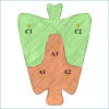
OLGA sample protocol for the gastritis staging.

Similar articles

Cited by

References

    1. Van Zanten SJ, Dixon MF, Lee A. The gastric transitional zones: neglected links between gastroduodenal pathology and helicobacter ecology. Gastroenterology 1999;116:1217-29. https://doi.org/10.1016/s0016-5085(99)70025-9 10.1016/s0016-5085(99)70025-9 - DOI - PubMed
    1. Hunt RH, Camilleri M, Crowe SE, et al. . The stomach in health and disease. Gut 2015;64:1650-68. https://doi.org/10.1136/gutjnl-2014-307595 10.1136/gutjnl-2014-307595 - DOI - PMC - PubMed
    1. Marshall BJ, Warren JR. Unidentified curved bacilli in the stomach of patients with gastritis and peptic ulceration. Lancet. 1984;1(8390):1311-5. https://doi.org/10.1016/s0140-6736(84)91816-6 10.1016/s0140-6736(84)91816-6 - DOI - PubMed
    1. Schistosomes liver flukes and Helicobacter pylori. IARC Working Group on the Evaluation of Carcinogenic Risks to Humans. Lyon, 7-14 June 1994. IARC Monogr Eval Carcinog Risks Hum 1994;61:1-241. - PMC - PubMed
    1. Okuda M, Sugiyama T, Fukunaga K, et al. . A strain-specific antigen in Japanese Helicobacter pylori recognized in sera of Japanese children. Clin Diagn Lab Immunol 2005;12:1280-4. https://doi.org/10.1128/CDLI.12.11.1280-1284.2005 10.1128/CDLI.12.11.1280-1284.2005 - DOI - PMC - PubMed

LinkOut - more resources