Review
. 2020 Sep;112(3):166-185.
doi: 10.32074/1591-951X-166.

Precancerous lesions of the stomach, gastric cancer and hereditary gastric cancer syndromes

Affiliations
Review

Precancerous lesions of the stomach, gastric cancer and hereditary gastric cancer syndromes

Irene Gullo et al. Pathologica. 2020 Sep.

Abstract

Gastric cancer accounts for about 6% of cancers worldwide, being the fifth most frequently diagnosed malignancy and the third leading cause of cancer related death. Gastric carcinogenesis is a multistep and multifactorial process and is the result of the complex interplay between genetic susceptibility and environmental factors. The identification of predisposing conditions and of precancerous lesions is the basis for screening programs and early stage treatment. Furthermore, although most gastric cancers are sporadic, familial clustering is observed in up to 10% of patients. Among them, hereditary cases, related to known cancer susceptibility syndromes and/or genetic causes are thought to account for 1-3% of all gastric cancers. The pathology report of gastric resections specimens therefore requires a standardized approach as well as in depth knowledge of prognostic and treatment associated factors.

Keywords: gastric adenocarcinoma; gastric cancer; gastric dysplasia; hereditary diffuse gastric cancer (HDGC); hereditary gastric cancer syndromes.


Conflict of interest statement

Conflict of interest

The Authors declare no conflict of interest.

Figures
Figure 1.
Figure 1.
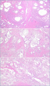
Two examples of intramucosal gastric carcinoma. In contrast to high grade dysplasia, intramucosal carcinoma shows marked glandular crowding and cribriform pattern (upper image, HE, magnification 10x). The bottom image shows a very well differentiated intramucosal carcinoma with crawling pattern (HE, magnification 10x).
Figure 2.
Figure 2.
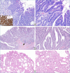
Gastric adenomas: (a) foveolar type adenoma with low grade dysplasia (HE, left image, magnification 40x, right image, magnification 10x) showing diffuse immunoreactivity for MUC5AC (inset); (b) intestinal type adenoma associated to mucinous carcinoma invading the submucosa (arrow) (HE, left image, magnification 4x); the image on the right represents an area of intestinal-type low grade dysplasia with tubular/villous morphology (HE, magnification 10x); (c) pyloric gland adenoma (HE, left image, magnification 4x, right image, magnification 20x).
Figure 3.
Figure 3.
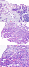
Hyperplastic polyp with dysplasia (HE, upper image 4x) and intramucosal carcinoma (HE, middle image, magnification 40x, bottom image, magnification 10x).
Figure 4.
Figure 4.
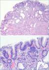
Fundic gland polyp with focus of low grade, foveolar-type dysplasia (upper image, HE, magnification 4x; (upper image, HE, magnification 20x). The patient had attenuated variant of familial adenomatous polyposis.
Figure 5.
Figure 5.
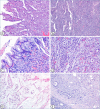
Main histopathological subtypes of gastric cancer: (a) papillary and tubulo-papillary gastric adenocarcinoma (HE, magnification 10x); (b) tubular adenocarcinoma with solid (high grade) areas (HE, magnification 10x); (c) poorly cohesive gastric cancer of the signet ring cell type (HE, magnification 20x); this tiny intramucosal focus was found in a prophylactic gastrectomy specimen in a CDH1 variant carrier; (d) poorly cohesive gastric cancer not otherwise specified (HE, magnification 20x); in this case the poorly cohesive cells show pleomorphic and plasmacytoid features; (e) mucinous adenocarcinoma, with and signet ring cells floating in mucin lakes (HE, magnification 20x); (f) mixed gastric cancer (HE, magnification 20x).
Figure 6.
Figure 6.
Rare histopathological variant of gastric cancer: (a) gastric cancer with lymphoid stroma showing abundant lymphoplasmacytic infiltrate (HE, magnification 10x); this case was associated to EBV infection, as evaluated by EBER-in situ hybridization (inset); (b) hepatoid gastric carcinoma with numerous hyaline globules (HE, magnification 20x); (c) micropapillary gastric carcinoma, with artefactual spaces at the periphery of the nests and inverted cell polarity (HE, 20x); (d) adenosquamous gastric carcinoma (HE, magnification 20x).
Figure 7.
Figure 7.
Gastric cancer arising in a stomach with gastritis cystica profunda: in gastritis cystica profunda gastric epithelium is displaced into the gastric wall (upper image, HE, magnification 4x); note the presence of lamina propria-like stroma surrounding the cystically dilated gastric glands; this case was associated to well differentiated tubular gastric adenocarcinoma (middle image, HE, 10x) with a mucinous component (bottom image, HE, magnification 10x).

Similar articles

Cited by

References

    1. Bray F, Ferlay J, Soerjomataram I, et al. . Global cancer statistics 2018: Globocan estimates of incidence and mortality worldwide for 36 cancers in 185 countries. CA Cancer J Clin 2018;68:394-424. https://doi.org/10.3322/caac.21492 10.3322/caac.21492 - DOI - PubMed
    1. Gullo I, Carneiro F, Oliveira C, et al. . Heterogeneity in gastric cancer: From pure morphology to molecular classifications. Pathobiology 2018;85:50-63. https://doi.org/10.1159/000473881 10.1159/000473881 - DOI - PubMed
    1. Eusebi LH, Telese A, Marasco G, et al. . Gastric cancer prevention strategies: A global perspective. J Gastroenterol Hepatol 2020. Online ahead of print. https://doi.org/10.1111/jgh.15037 10.1111/jgh.15037 - DOI - PubMed
    1. Fukayama M, Abe H, Kunita A, et al. . Thirty years of epstein-barr virus-associated gastric carcinoma. Virchows Arch 2020;476;353-5. https://doi.org/10.1007/s00428-019-02724-4 10.1007/s00428-019-02724-4 - DOI - PubMed
    1. Sanduleanu S, Jonkers D, De Bruine A, et al. . Non-helicobacter pylori bacterial flora during acid-suppressive therapy: differential findings in gastric juice and gastric mucosa. Aliment Pharmacol Ther 2001;15;379-88. https://doi.org/10.1046/j.1365-2036.2001.00888.x 10.1046/j.1365-2036.2001.00888.x - DOI - PubMed

MeSH terms