

Celiac disease: histology-differential diagnosis-complications. A practical approach
- PMID: 33179621
- PMCID: PMC7931573
- DOI: 10.32074/1591-951X-157
Celiac disease: histology-differential diagnosis-complications. A practical approach
Abstract
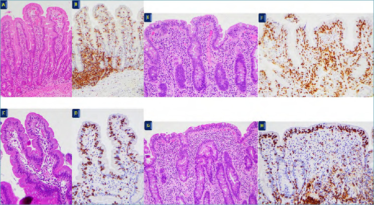
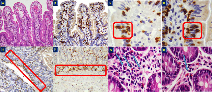
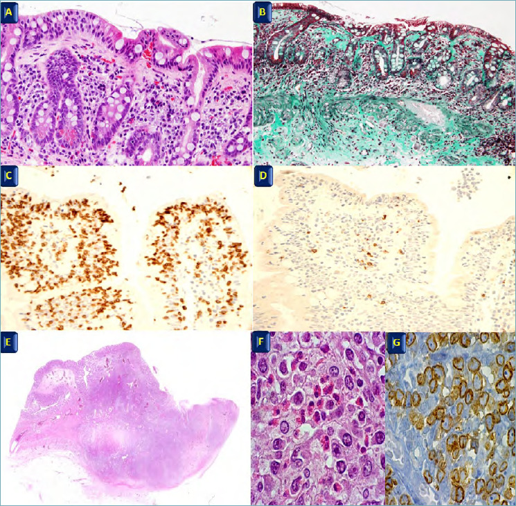
Celiac disease is a multi-factorial chronic inflammatory intestinal disease, characterized by malabsorption resulting from mucosal injury after ingestion of wheat gluten or related rye and barley proteins. Inappropriate T-cell-mediated immune response against ingested gluten in genetically predisposed people, leads to characteristic histological lesions, as villous atrophy and intraepithelial lymphocytosis. Nevertheless, celiac disease is a comprehensive diagnosis with clinical, serological and genetic characteristics integrated with histological features. Biopsy of duodenal mucosa remains the gold standard in the diagnosis of celiac disease with the recognition of the spectrum of histological changes and classification of mucosa damage based on updated Corazza-Villanacci system. Appropriate differential diagnosis evaluation and clinical context also for the diagnosis of complications is, moreover, needed for correct histological features interpretation and clinical management.
Keywords: celiac disease; gluten; small bowel; sprue.
Copyright © 2020 Società Italiana di Anatomia Patologica e Citopatologia Diagnostica, Divisione Italiana della International Academy of Pathology.
Conflict of interest statement
The Authors declare no conflict of interest.
Figures
References
-
- Robert ME, Crowe SE, Burgart L, et al. . Statement on best practices in the use of pathology as a diagnostic tool for celiac disease: a guide for clinicians and pathologists. Am J Surg Pathol 2018;42:e44-e58. https://doi.org/10.1097/PAS.0000000000001107 10.1097/PAS.0000000000001107 - DOI - PubMed
-
- Volta U, Caio G, Stanghellini V, et al. . Non-celiac gluten sensitivity: questions still to be answered despite increased awarness. Cell Mol Immunol 2013;10:383-92. https://doi.org/10.1038/cmi.2013.28 10.1038/cmi.2013.28 - DOI - PMC - PubMed
-
- Caio G, Volta U, Sapone A, et al. . Celiac disease: a comprehensive current review. BMC Medicine 2019;17:142-62. https://doi.org/10.1186/s12916-019-1380-z 10.1186/s12916-019-1380-z - DOI - PMC - PubMed
-
- Ludvigsson JF, Rubio-Tapia A, van Dyke CT, et al. . Increasing incidence of celiac disease in a North American population. Am J Gastroenterol 2013:108:818-24. https://doi.org/10.1038/ajg.2013.60 10.1038/ajg.2013.60 - DOI - PMC - PubMed
-
- Mustalahti K, Catassi C, Reunanen A, et al. . The prevalence of celiac disease in Europe: results of a centralized, international mass screening project. Ann Med 2010;42:587-95. https://doi.org/10.3109/07853890.2010.505931 10.3109/07853890.2010.505931 - DOI - PubMed
Publication types
MeSH terms
Substances
LinkOut - more resources
Full Text Sources
Medical
