Review
. 2021 Feb;56(1):1-16.
doi: 10.1080/10409238.2020.1832438. Epub 2020 Nov 12.

Human de novo purine biosynthesis

Affiliations
Review

Human de novo purine biosynthesis

Vidhi Pareek et al. Crit Rev Biochem Mol Biol. 2021 Feb.

Abstract

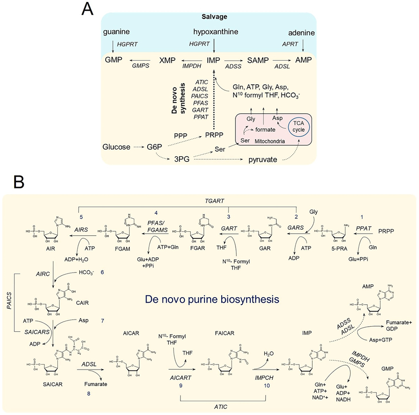
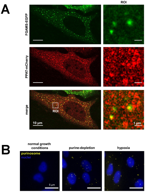
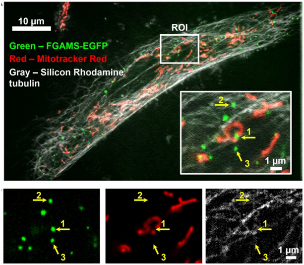
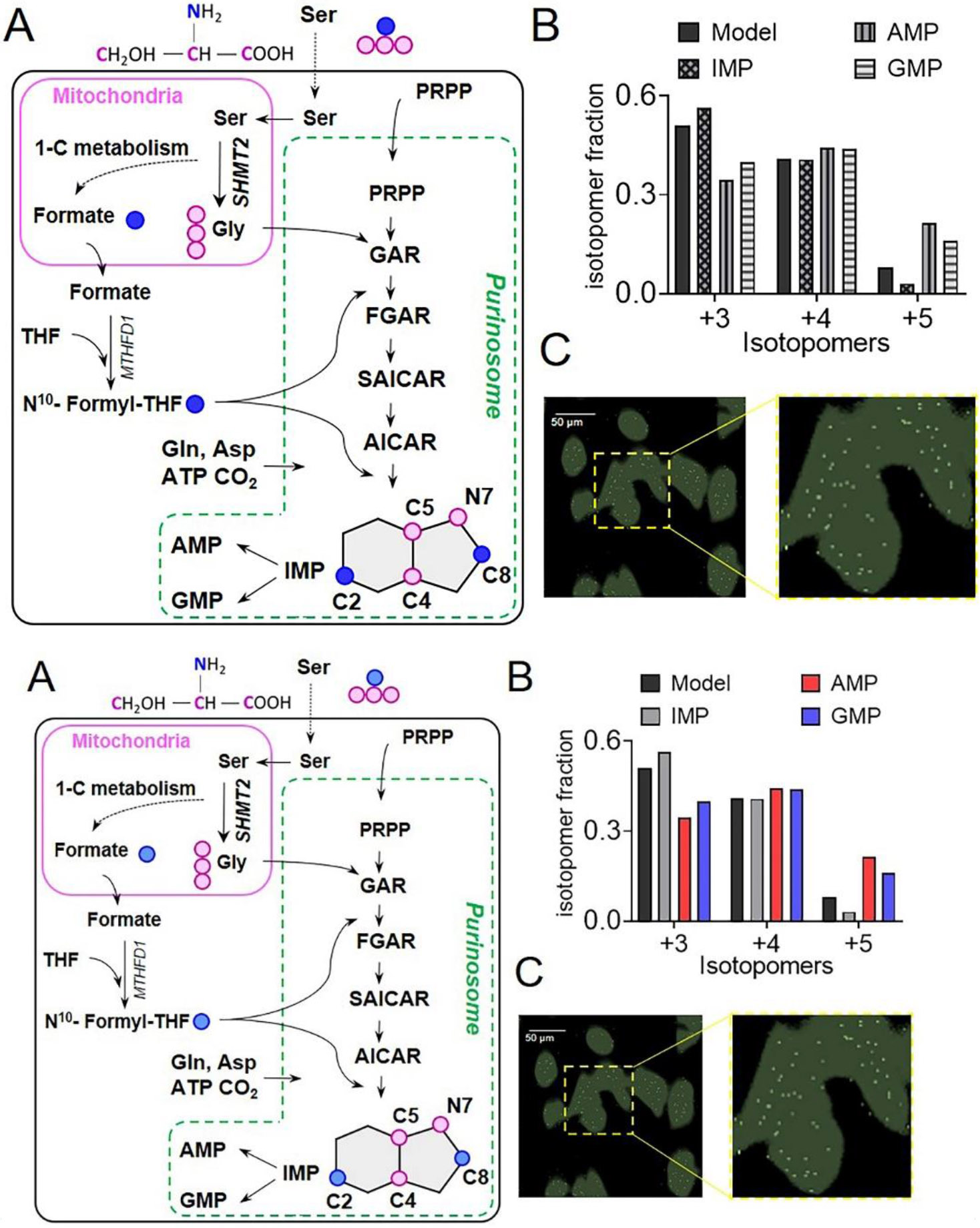
The focus of this review is the human de novo purine biosynthetic pathway. The pathway enzymes are enumerated, as well as the reactions they catalyze and their physical properties. Early literature evidence suggested that they might assemble into a multi-enzyme complex called a metabolon. The finding that fluorescently-tagged chimeras of the pathway enzymes form discrete puncta, now called purinosomes, is further elaborated in this review to include: a discussion of their assembly; the role of ancillary proteins; their locus at the microtubule/mitochondria interface; the elucidation that at endogenous levels, purinosomes function to channel intermediates from phosphoribosyl pyrophosphate to AMP and GMP; and the evidence for the purinosomes to exist as a protein condensate. The review concludes with a consideration of probable signaling pathways that might promote the assembly and disassembly of the purinosome, in particular the identification of candidate kinases given the extensive phosphorylation of the enzymes. These collective findings substantiate our current view of the de novo purine biosynthetic metabolon whose properties will be representative of how other metabolic pathways might be organized for their function.

Keywords: metabolism de novo purine biosynthesis purinosome metabolon substrate channeling condensate signaling.


Conflict of interest statement

DISCLOSURE STATEMENT

No potential conflict of interest was reported by the authors.

Figures

Figure 1.
Figure 1.. Purine salvage and de novo purine biosynthesis (DNPB).
(A) Purines are regenerated either by salvage from the bases hypoxanthine, guanine, and adenine; or synthesized de novo from PRPP, utilizing building block substrates generated by other metabolic processes. Glucose metabolism generates- 1) Glucose-6-phosphate (G6P) that can generate PRPP via the pentose phosphate pathway (PPP); 2) 3-phosphoglycerate (3PG) that can generate Ser via the serine biosynthesis pathway; and 3) pyruvate that fuels the TCA cycle inside mitochondria, which in turn maintains a stable supply of aspartic acid. Conversion of Ser to Gly and formate is carried out inside mitochondria and requires the involvement of one carbon metabolism enzymes. Enzyme names are represented in italicized letters. (B) Schematic shows the step by step conversion of PRPP to AMP and GMP constituting the DNPB pathway. A cascade of 10 reactions catalyzed by the six DNPB enzymes produces IMP, which is either converted to AMP or GMP in two additional each way. It is an energy intensive pathway that requires three amino acids- Gly, Asp, Gln- and the cofactor N10-formyl tetrahydrofolate (N10- formyl THF). All the purine intermediate and enzyme acronym expansions are defined in the text.
Figure 2.
Figure 2.. Detection of Purinosomes on the Overexpressed and Endogenous Protein Levels.
(A) A purine-depleted HeLa cell showing the colocalization of FGAMS-EGFP (green) and PPAT-mCherry (red) identifying purinosomes (yellow puncta) in a transient, overexpressed system. The image was modified from Pedley and Benkovic (Pedley and Benkovic, 2018). (B) The colocalization of endogenous FGAMS and ADSL by proximity ligation assays to identify purinosomes (yellow puncta) under purine-depleted and hypoxic growth conditions. The image was modified from the research originally published in the Journal of Biological Chemistry (Doigneaux et al., 2020) and used with permission from C. Doigneaux and A. Tavassoli.
Figure 3.
Figure 3.. Interplay of Purinosomes with Mitochondria and Microtubules.
Colocalization of purinosomes (FGAMS-EGFP, green), mitochondria (Mitotracker Red, red), and microtubules (silicon rhodamine tubulin, gray) in an HPRT-deficient fibroblast by high-resolution confocal microscopy (VT-iSIM). Inset shows a magnified region of interest (ROI) with individual purinosome colocalizations annotated with respect to the mitochondrion (1), microtubule (2), or neither subcellular structure-of-interest (3). Individual channels for purinosomes, mitochondria, and microtubules of the ROI are shown underneath. Image was modified from Chan et al., 2018 (Chan et al., 2018).
Figure 4.
Figure 4.. HeLa cells show channeled DNPB.
(A) In three separate steps upstream of IMP, 13C3, 15N Ser derived labeled 13C2, 15N Gly and 13C formate get incorporated into the intermediates and the end-product purines synthesized by DNPB, allowing monitoring of cellular DNPB flux. The unique isotopomer species in IMP, AMP, and GMP- +3, +4, and +5- all have one 13C2, 15N Gly; and 0, 1 or 2 13C formate incorporations, respectively. For clarity, only the relevant steps and intermediates are shown. Blue circles: 13C labeled β-carbon of Ser; pink circles: 13C, 15N labeled Ser backbone atoms. (B) The fractional abundance of the de novo synthesized purine isotopomers, +3, +4, and +5 can be predicted assuming uniform cytosolic abundance of the labeled substrates; a similar distribution of the three isotopomers is to be expected for IMP and all the downstream purines, including AMP and GMP. While the isotopomeric distribution of IMP closely resembles the model, AMP and GMP show a different isotopomeric distribution. The underlying reason being a higher labeled Gly and formate enrichment in AMP and GMP compared to that predicted by the model. Data from one representative experiment are shown. (C) A representative GCIB-SIMS image of purine depleted HeLa cells grown in 15N Ser supplemented medium to allow 15N label incorporation in purines. Overlay of the pixels with high 15N AICAR abundance (white) and cell image generated using total ion current (green) is shown. A zoomed-in view shows the spatial distribution of AICAR ‘metabolic hotspots’ in a region of interest highlighted with yellow box in (C). Figures are prepared from the data originally reported and presented in (Pareek et al., 2020).
Figure 5.
Figure 5.. The Regulation of de novo Purine Biosynthesis and Purinosome Formation.
(A) Pathway activation and/or purinosome formation has been associated with signaling through the GPCR, MAPK, and PI3K/AKT signaling pathways. Downstream of AKT is mTOR, whose activity assists in the colocalization of purinosomes with mitochondria. Dashed lines suggest an association or correlation, whereas solid lines represent a direct interaction or consequence. (B) The phosphorylation sites identified on PFAS/FGAMS as determined our recent post-translational modification study (Liu et al., 2019) and other global phosphoproteomic studies recorded on PhosphoSite. Solid red circles and white circles represent those phosphorylation events identified under purine supplemented and purine-depleted growth conditions, respectively. Red stripped circles were either present in both conditions or the site preference is unknown. Kinase predictions are a combination from ScanSite and/or Liu et al., 2019.
Figure 6.
Figure 6.. A model of purinosomes, the DNPB metabolon.
Results from high resolution fluorescence imaging, biochemical, proteomic, metabolomic, and chemical imaging studies taken together suggest that purinosomes are constituted by at least nine enzyme and plausibly more ancillary proteins that assist in stabilization and dynamics of the complex. The HSP90/70 machinery and signaling pathways involving kinases and other post translationally modifying enzymes regulate the process of assembly of an active purinosome. A majority of purinosomes reside proximal to mitochondria-microtubule junction and act as cellular metabolic hot-spots that synthesize AMP and GMP in a highly channeled manner. Purinosome assisted AMP+GMP synthesis has ~7 times higher flux than the diffusive synthesis of IMP.

Similar articles

Cited by

References

    1. Lane AN, Fan TW. (2015).Regulation of mammalian nucleotide metabolism and biosynthesis. Nucleic Acids Res, 43, 2466–85 - PMC - PubMed
    1. Pedley AM, Benkovic SJ. (2017).A New View into the Regulation of Purine Metabolism: The Purinosome. Trends Biochem Sci, 42, 141–54 - PMC - PubMed
    1. Garcia-Gil M, Camici M, Allegrini S, Pesi R, Petrotto E, Tozzi MG. (2018).Emerging Role of Purine Metabolizing Enzymes in Brain Function and Tumors. Int J Mol Sci, 19 - PMC - PubMed
    1. Jinnah HA. (2009).Lesch-Nyhan disease: from mechanism to model and back again. Dis Model Mech, 2, 116–21 - PMC - PubMed
    1. Jurecka A, Zikanova M, Kmoch S, Tylki-Szymanska A. (2015).Adenylosuccinate lyase deficiency. J Inherit Metab Dis, 38, 231–42 - PMC - PubMed

Publication types

MeSH terms

LinkOut - more resources