. 2020 Nov 7;12(21):21758-21776.
doi: 10.18632/aging.103984. Epub 2020 Nov 7.

Caspase-3 knockout attenuates radiation-induced tumor repopulation via impairing the ATM/p53/Cox-2/PGE2 pathway in non-small cell lung cancer

Affiliations

Caspase-3 knockout attenuates radiation-induced tumor repopulation via impairing the ATM/p53/Cox-2/PGE2 pathway in non-small cell lung cancer

Minghui Zhao et al. Aging (Albany NY). .

Abstract

Radiotherapy is an effective treatment for non-small cell lung cancer (NSCLC). However, irradiated, dying tumor cells generate potent growth stimulatory signals during radiotherapy that promote the repopulation of adjacent surviving tumor cells to cause tumor recurrence. We investigated the function of caspase-3 in NSCLC repopulation after radiotherapy. We found that radiotherapy induced a DNA damage response (DDR), activated caspase-3, and promoted tumor repopulation in NSCLC cells. Unexpectedly, caspase-3 knockout attenuated the ataxia-telangiectasia mutated (ATM)/p53-initiated DDR by decreasing nuclear migration of endonuclease G (EndoG), thereby reducing the growth-promoting effect of irradiated, dying tumor cells. We also identified p53 as a regulator of the Cox-2/PGE2 axis and its involvement in caspase-3-induced tumor repopulation after radiotherapy. In addition, injection of caspase-3 knockout NSCLC cells impaired tumor growth in a nude mouse model. Our findings reveal that caspase-3 promotes tumor repopulation in NSCLC cells by activating DDR and the downstream Cox-2/PGE2 axis. Thus, caspase-3-induced ATM/p53/Cox-2/PGE2 signaling pathway could provide potential therapeutic targets to reduce NSCLC recurrence after radiotherapy.

Keywords: DNA damage response; caspase-3; non-small cell lung cancer; radiotherapy; tumor repopulation.


Conflict of interest statement

CONFLICTS OF INTEREST: The authors declare no conflicts of interest.

Figures

Figure 1
Figure 1
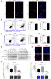
Radiations induce DNA damage, caspase-3 activation, and tumor repopulation in NSCLC cells. (A) Confocal images of immunostained A549 and H460 cells showing γH2AX foci following 8 Gy irradiation at 48 h. Scale bars: 25 μm. (B, C) The left panel shows flow cytometry analysis of A549 (B) and H460 (C) cell death after 0 Gy or 8 Gy irradiation on day 3. Apoptosis was monitored by Annexin V/propidium iodide (PI) double staining. The right panel shows quantitative analysis of early apoptosis and total cell death in 0 Gy- or 8 Gy-irradiated A549 (B) and H460 (C) cells (***p<0.001, Student’s t test, n = 3). (D) Cleaved caspase-3 induced by 8 Gy radiations was assayed by western blotting, and β-tubulin and glyceraldehyde 3-phosphate dehydrogenase (GAPDH) served as loading controls. (E) Representative confocal images of immunostained A549 and H460 cells showing cleaved caspase-3 following exposure to 8 Gy radiations on day 3. Scale bars: 25 μm. (F) The 8 Gy-irradiated NSCLC cells promoted the growth of living NSCLC reporter cells. The upper panel depicts luciferase activities showing the growth of A549 Fluc and H460 Fluc cells that were seeded alone or with 0 Gy- or 8 Gy-irradiated NSCLC cells. The lower panel shows the representative bioluminescence images (**p<0.01, ***p<0.001, one-way analysis of variance [ANOVA], n = 4).
Figure 2
Figure 2
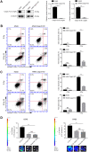
Casp3 KO attenuates radiation-induced apoptosis and growth-promoting effect of dying NSCLC cells. (A) Western blot analysis of the expression of caspase-3 in Casp3 KO A549 and H460 cells generated using the CRISPR/Cas9 system. β-tubulin was used as the loading control (***p<0.001, Student’s t test, n = 3). (B, C) The left panel shows the flow cytometry analysis of cell death in A549 and A549/Casp3 KO (B) and H460 and H460/Casp3 KO (C) cells following irradiation. Apoptotic cells were analyzed by Annexin V/propidium iodide (PI) double staining. The right panel shows the quantitative analysis of early apoptosis and total cell death in 0 Gy- or 8 Gy-irradiated control and A549/Casp3 KO (B) and H460/Casp3 KO (C) cells (***p<0.001, NS = not significant, Student’s t test, n = 3). (D) Casp3 KO significantly decreased the growth-promoting effect of 8 Gy-irradiated NSCLC cells on living NSCLC reporter cells. The upper panel depicts the luciferase activities showing the growth of A549 Fluc or H460 Fluc cells that were seeded with 8 Gy-irradiated wild-type or Casp3 KO cells or alone. The lower panel shows the representative bioluminescence images (***p<0.001, NS = not significant, one-way analysis of variance [ANOVA], n = 4).
Figure 3
Figure 3
Caspase-3-dependent PGE2 production in dying NSCLC cells induces tumor repopulation. (A) Western blot analysis of Cox-2 levels at various time intervals after 8 Gy irradiation of wild-type and Casp3 KO NSCLC cells (***p<0.001, one-way analysis of variance [ANOVA], n = 3). (B) Quantitative polymerase chain reaction (qPCR) analysis of Cox-2 in wild-type and Casp3 KO NSCLC cells at indicated times after 8 Gy irradiation (*p<0.05, **p<0.01, ***p<0.001, one-way ANOVA, n = 3). (C) Levels of prostaglandin E2 (PGE2) in culture supernatants of wild-type and Casp3 KO NSCLC cells at 48 h after 8 Gy irradiation were measured using enzyme-linked immunosorbent assay (ELISA) (***p<0.001, one-way ANOVA, n = 3). (D) A selective Cox-2 inhibitor, celecoxib, abrogated the pro-proliferation effects of dying NSCLC cells on Fluc cells in a dose-dependent manner (*p<0.05, ***p<0.001, NS = not significant, one-way ANOVA, n = 4).
Figure 4
Figure 4
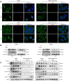
Casp3 KO attenuates the DDR via ATM/p53 signaling in irradiated NSCLC cells. (A) Immunofluorescence analysis of 8 Gy-irradiated wild-type and Casp3 KO NSCLC cells co-stained for EndoG and γH2AX foci at 48 h. Scale bars: 25 μm. (B) Western blot analysis of EndoG in the cytoplasmic and nuclear fractions of 8 Gy-irradiated wild-type and Casp3 KO NSCLC cells at 48 h. β-tubulin and Histone H3 were used as cytoplasmic and nuclear loading controls, respectively. (C) Levels of DNA damage response (DDR)-related proteins ATM, pATM (S1981), Chk2, pChk2 (T68), p53, and pp53 (S15) were measured by western blotting at indicated times after 8 Gy irradiation of wild-type and Casp3 KO NSCLC cells. GAPDH was used as the loading control.
Figure 5
Figure 5
p53 induces Cox-2 in NSCLC cells. (A) Schematic representation of the luciferase reporter plasmid with the wild-type PTGS2 promoter sequence (PTGS2-WT) or mutant sequence (PTGS2-Mut). (B) A p53-dependent stimulation of PTGS2 promoter activity was demonstrated by luciferase assay. The 293T cells were co-transfected with p53 overexpression plasmid and PTGS2-WT plasmid, PTGS2-Mut plasmid, or vector alone. The pGMR-TK reporter was used as an internal transfection standard (***p<0.001, one-way analysis of variance [ANOVA], n = 3). (C, D) Quantitative polymerase chain reaction (qPCR) and western blot analysis showed that the mRNA and protein levels of Cox-2 were elevated by overexpression of p53 in wild-type and Casp3 KO NSCLC cells. Total RNA and proteins were extracted after transfection for 24 h and 48 h, respectively (***p<0.001, Student’s t test, n = 3).
Figure 6
Figure 6
Casp3 KO inhibits tumor formation, and radiations activate the ATM/p53/Cox-2 axis in vivo. (A) Treatment scheme for nude mice. (B) Images of tumors obtained on day 22. (C) The tumor volume of xenografts was measured with calipers every 2 or 3 days (**p<0.01, ***p<0.001, Student’s t test, n = 7). (D) Representative photomicrographs of hematoxylin and eosin (H&E) and immunohistochemical staining of caspase-3, cleaved caspase-3, ATM, pATM (S1981), Chk2, pChk2 (T68), p53, pp53 (S15), and Cox-2 in tumor tissues. Scale bars: 50 μm.
Figure 7
Figure 7
Schematic illustration of the proposed mechanism of radiation-induced tumor repopulation in NSCLC. Radiation-induced DNA double-strand breaks (DSBs) activate the DNA damage response (DDR) and caspase-3. Activated caspase-3 regulates the EndoG nuclear translocation and thus participates in the DDR by regulating ATM/p53 signaling, which activates the Cox-2/PGE2 axis in dying NSCLC cells, consequently enhancing the proliferation of living tumor cells.

Similar articles

Cited by

References

    1. Jemal A, Siegel R, Ward E, Hao Y, Xu J, Thun MJ. Cancer statistics, 2009. CA Cancer J Clin. 2009; 59:225–49. 10.3322/caac.20006 - DOI - PubMed
    1. Herbst RS, Heymach JV, Lippman SM. Lung cancer. N Engl J Med. 2008; 359:1367–80. 10.1056/NEJMra0802714 - DOI - PMC - PubMed
    1. Dağoğlu N, Karaman Ş, Arifoğlu A, Küçücük S, Oral EN. Definitive radiotherapy in locally advanced non-small cell lung cancer: dose and fractionation. Balkan Med J. 2014; 31:278–85. 10.5152/balkanmedj.2014.14496 - DOI - PMC - PubMed
    1. Withers HR, Taylor JM, Maciejewski B. The hazard of accelerated tumor clonogen repopulation during radiotherapy. Acta Oncol. 1988; 27:131–46. 10.3109/02841868809090333 - DOI - PubMed
    1. Kim JJ, Tannock IF. Repopulation of cancer cells during therapy: an important cause of treatment failure. Nat Rev Cancer. 2005; 5:516–25. 10.1038/nrc1650 - DOI - PubMed

Publication types

MeSH terms