. 2020 Nov 12;15(11):e0242337.
doi: 10.1371/journal.pone.0242337. eCollection 2020.

In-situ proliferation contributes to the accumulation of myeloid cells in the spleen during progressive experimental visceral leishmaniasis

Affiliations

In-situ proliferation contributes to the accumulation of myeloid cells in the spleen during progressive experimental visceral leishmaniasis

E Yaneth Osorio et al. PLoS One. .

Abstract

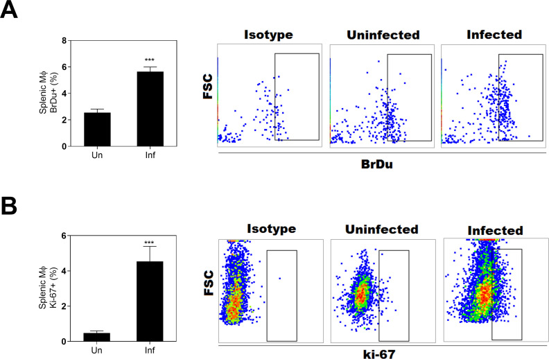
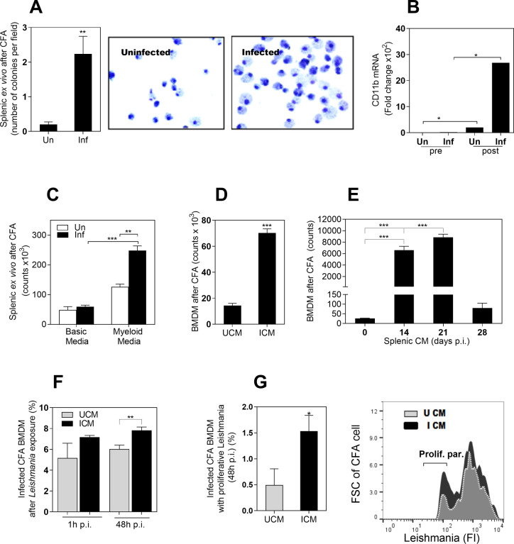
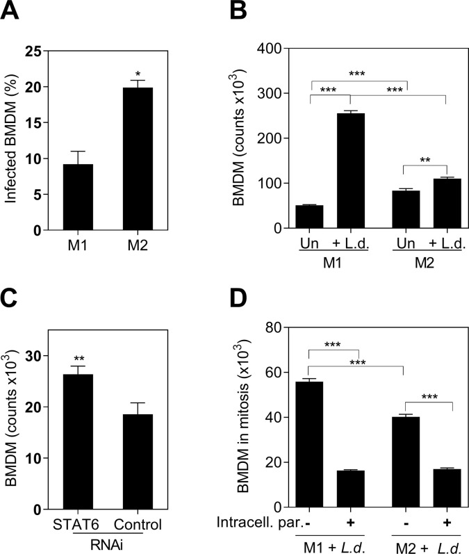
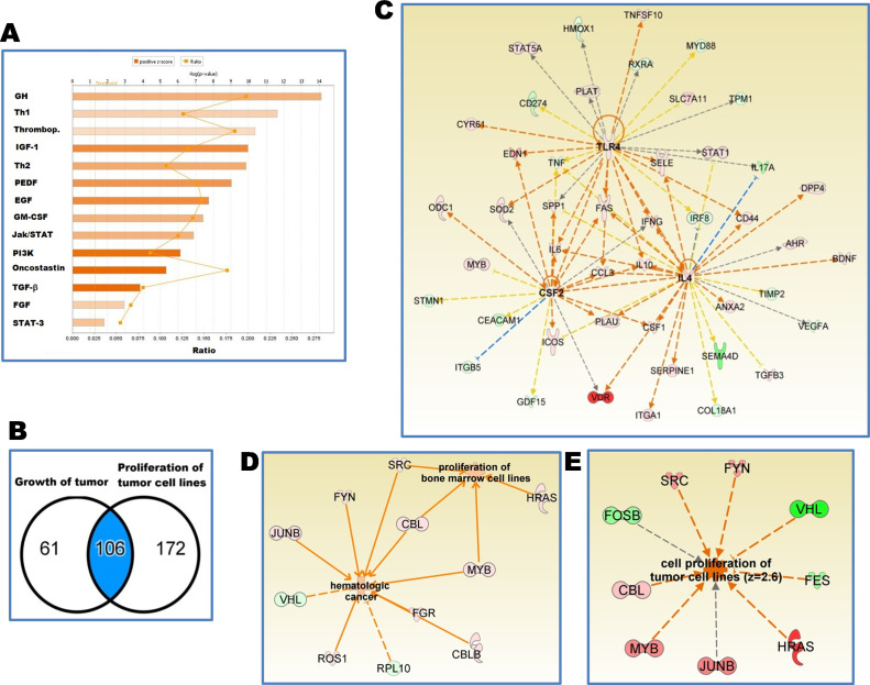
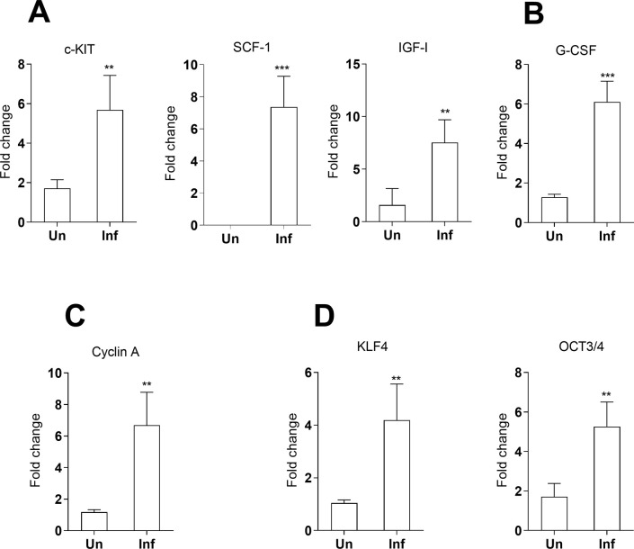
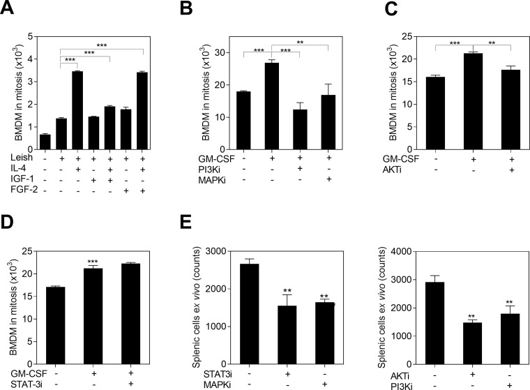
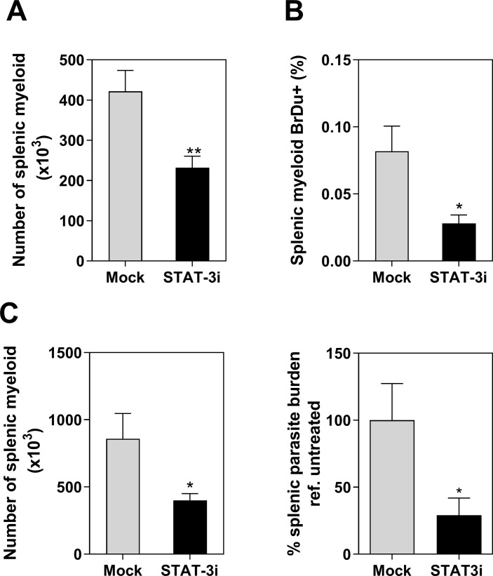
Visceral leishmaniasis (VL) is characterized by expansion of myeloid cells in the liver and spleen, which leads to a severe splenomegaly associated with higher risk of mortality. This increased cellularity is thought to be a consequence of recruitment of cells to the viscera. We studied whether the local proliferation of splenic myeloid cells contributes to increased splenic cellularity. We found that a monocyte-like population of adherent splenic cells from Leishmania donovani-infected hamsters had enhanced replicative capacity ex vivo and in vivo (BrdU incorporation, p<0.0001). In vitro assays demonstrated that proliferation was more pronounced in the proinflammatory M1 environment and that intracellular infection prevented proliferation. Secondary analysis of the published splenic transcriptome in the hamster model of progressive VL revealed a gene expression signature that included division of tumoral cells (Z = 2.0), cell cycle progression (Z = 2.3), hematopoiesis (Z = 2.8), proliferation of stem cells (Z = 2.5) and overexpression of proto-oncogenes. Regulators of myeloid cell proliferation were predicted in-silico (CSF2, TLR4, IFNG, IL-6, IL-4, RTK signaling, and STAT3). The in-silico prediction was confirmed with chemical inhibitors of PI3K/AKT, MAPK and STAT3 which decreased splenic myeloid cell division ex vivo. Hamsters infected with L. donovani treated with a STAT3 inhibitor had reduced in situ splenic myeloid proliferation (p = 0.03) and parasite burden. We conclude that monocyte-like myeloid cells have increased STAT3-dependent proliferation in the spleen of hamsters with visceral leishmaniasis and that inhibition of STAT3 reduces myeloid cell proliferation and parasite burden.


Conflict of interest statement

The authors have declared that no competing interests exist.

Figures

Fig 1
Fig 1. Splenic myeloid cells from hamsters infected with L. donovani are proliferative.
A, Proportion of splenic myeloid cells BrdU+ (DNA synthesis) in uninfected (Un) or infected hamsters (Inf) at 28 d p.i., (*** p<0.0001, Unpaired T-test) and representative flow cytometry dot-plot showing the BrdU+ cells. B, Proportion of splenic myeloid cells in mitosis (Ki-67+), at 28d p.i., (*** p<0.0001, Mann-Whitney Test) and representative flow cytometry dot-plot showing the proportion of ki-67+ cells.
Fig 2
Fig 2. The infected environment in VL induces the proliferation of myeloid cells.
A, Number of colonies in a colony formation assay (CFA) using splenic myeloid cells from uninfected hamsters (Un) or from hamsters infected with L. donovani (Inf). Colonies were counted on the inverted microscope after 12 days of incubation (**p<0.008, Mann-Whitney Test); Right, representative Giemsa staining of cells at day 12 of CFA, showing some bi-nucleated cells; B, Expression of the pan-myeloid CD11b marker before culture (pre) and at 12 days of CFA (post), determined by qPCR (*p<0.05, Kruskal-Wallis Test); C, Number of cells at 12 days of CFA with splenic myeloid cells exposed to a basic medium (without myeloid growth factors) or complete medium (with myeloid growth factors). The number of cells was determined by Cell Titer Glo (**p<0.01; ***p<0.0001, Kruskal-Wallis Test); D, Number of bone marrow derived cells (BMDM) at 12 days of exposure to a CFA with CM from spleen of uninfected (UCM) or infected hamsters (ICM) (***p<0.0001, Unpaired T test); E, Number of BMDM at 12 days of CFA exposed to splenic CM obtained from spleens of uninfected hamsters (time 0) or from hamsters infected with L. donovani at the indicated times post-infection (14–28 days p.i.) ***p<0.001, Tukey-Kramer Multiple Comparisons Test); F, Percentage of infected CFA cells after in vitro exposure to L. donovani promastigotes (1h or 48h of in vitro infection). CFA cells obtained after 12 days of BMDM exposed to UCM or ICM as determined by flow cytometry (**p = 0.03, Unpaired t test); G, Percentage of cells from CFA with proliferative parasites after 48h of in vitro infection with L. donovani promastigotes. Parasites were prelabeled with a fluorescent proliferation tracer before in vitro infection (*p< 0.004, Unpaired T test). Right, representative histogram showing fluorescence intensity of proliferative parasites (with diluted fluorescence) compared with parental parasites.
Fig 3
Fig 3. The proliferative capacity of BMDM was limited by intracellular L.donovani parasites and M2-polarization.
A, Proportion of infected M1 or M2 BMDM after 48 hours of exposure to L. donovani promastigotes (*p = 0.03, Unpaired t test, representative of 3 experiments); B, Absolute number of M1 or M2 BMDM stimulated with L. donovani promastigotes (+L.d.) or without parasite stimulation (Un). Number of cells counted by luminometry (*** p<0.001, **p<0.01, Tukey-Kramer Multiple Comparisons Test, representative of 3 different experiments); C, Number of BMDM after STAT6 silencing (STAT6 RNAi) compared with control silenced cells (control RNAi), 48h after exposure to L. donovani parasites (** p = 0.0079, Mann-Whitney Test). Data is the number of stimulated cells minus non-stimulated cells; D, Number of M1 or M2-polarized BMDM Ki-67+ (mitosis) containing intracellular parasites (+) or remaining free of intracellular parasites (-), after 48h of exposure to fluorescent-L. donovani (Number of cells in mitosis = percentage ki-67+ cells by flow cytometry x number of cells by luminometry/ 100) (*** p<0.0001, Tukey-Kramer Multiple Comparisons Test, representative of 3 different experiments).
Fig 4
Fig 4. Transcriptome of splenic myeloid cells from hamsters with VL show proliferative pathways characteristic of cancer diseases.
A, Signaling pathways associated with cell proliferation of tumor cell lines in a set of 278 differentially expressed genes identified in the transcriptome of splenic myeloid cells of hamsters with VL; B, Venn diagram representing a set of 106 differentially expressed genes associated with functions of proliferation of tumor cell lines and growth of tumor; C, Predicted upstream regulators of those 106 differentially expressed genes: Interleukin-4 (IL-4, Z = 2.32), Granulocyte-macrophage colony-stimulating factor = Colony stimulating factor 2 (CSF2, Z = 2.0) and Toll-like receptor 4 (TLR4, Z = 2.32); Orange arrows: lead to activation; blue arrows: lead to inhibition; yellow arrows: conflicting finding; grey arrows undetermined; pink to red gene: increased expression; green gene: decreased expression; D,E, Differentially expressed proto-oncogenes and tumor suppressor genes that lead to cell division of tumoral cells. Relationships and functions predicted with the IPA software (Z score > 2.0). Differential gene expression found in the hamster splenic myeloid transcriptome [17] with a FDR<0.05 and fold change 2.0 with reference to uninfected.
Fig 5
Fig 5. Expression of genes associated with stem cells in the spleen of hamsters with VL as determined by real time qPCR.
A-C, Genes expressed by splenic adherent myeloid cells; D, Genes expressed in total spleen of uninfected hamsters (Un) or hamsters infected with L. donovani (Inf) at 28d p.i. Data represents fold change with reference to uninfected (mean and SE of 3 experiments, 8 hamsters per group per experiment). c-KIT: p = 0.03; SCF: p = 0.0067, G-CSF: p = 0.0001; IGF-1: p = 0.0051; cyclin A: p = 0.0048; KLF4: p = 0.0064; OCT3/4: p = 0.02, Mann-Whitney Test.
Fig 6
Fig 6. The proliferation of myeloid cells exposed to L. donovani depends on PI3K/AKT, MAPK and STAT3 signaling.
A, Mitosis of BMDM (ki-67+) after 72h of in vitro exposure to L. donovani (Leish) and stimulation with IGF-1, FGF-2 or IL-4 (***p<0.001, Dunnett Multiple Comparisons Test); B, C, Mitosis of BMDM (ki-67+) after exposure to L. donovani, stimulation with GM-CSF, and treatment with either 1.5μM PI3-kinase inhibitor (LY29402), 10μM MAPK kinase inhibitor (PD98059), or 0.2uM AKT inhibitor (MK-2206) (***p<0.001, **p<0.01, Tukey-Kramer Multiple Comparisons Test); D, Mitosis of BMDM (ki-67+) after exposure to L. donovani, GM-CSF and 25μM STAT3 inhibitor (S31-201); Cells in mitosis = proportion of Ki-67+ cells by flow cytometry x number of cells/100; E, Number of splenic myeloid cells from infected hamsters after ex vivo treatment with the same concentrations of chemical inhibitors (***p<0.001, **p<0.01, Tukey-Kramer Multiple Comparisons Test, with reference to untreated cells).
Fig 7
Fig 7. STAT3 inhibitors modulate the proliferation of splenic myeloid cells in hamsters infected with L. donovani.
A, Number of splenic myeloid cells recovered from infected hamsters after treatment with STAT3 inhibitor (cucurbitacin) starting 14 days p.i.(light microscopy) (** p = 0.007, Unpaired t test); B, Proportion of in situ proliferative splenic myeloid cells from the same groups of hamsters injected i.p. with BrdU (BrdU pulse-chase incorporation, flow cytometry) (* p = 0.03, Unpaired t test); C, Number of myeloid cells and Percentage of splenic parasite burden in hamsters infected with L. donovani treated with the STAT3 inhibitor (Stattic) starting at 18–21 days p.i. Percentage of parasite burden in treated vs. mock treated hamsters (RT qPCR) (*p = 0.02, Mann Whitney test, n = 9–10 hamsters per group, 2 experiments).

Similar articles

Cited by

References

    1. Gallina V, Binazzi R, Golemi A, Farsad M, Weiss G, Wiedermann CJ. Imported visceral leishmaniasis—unexpected bone marrow diagnosis in a patient with fever, pancytopenia, and splenomegaly. Am J Blood Res. 2014;4(2):101–5. - PMC - PubMed
    1. Veress B, Omer A, Satir AA, El Hassan AM. Morphology of the spleen and lymph nodes in fatal visceral leishmaniasis. Immunology. 1977;33(5):605–10. - PMC - PubMed
    1. Zuckerman A. Current status of the immunology of blood and tissue Protozoa. I. Leishmania. Exp Parasitol. 1975;38(3):370–400. 10.1016/0014-4894(75)90123-x - DOI - PubMed
    1. Sarkari B, Naraki T, Ghatee MA, Abdolahi Khabisi S, Davami MH. Visceral Leishmaniasis in Southwestern Iran: A Retrospective Clinico-Hematological Analysis of 380 Consecutive Hospitalized Cases (1999–2014). PLoS One. 2016;11(3):e0150406 10.1371/journal.pone.0150406 - DOI - PMC - PubMed
    1. Abidin BM, Hammami A, Stäger S, Heinonen KM. Infection-adapted emergency hematopoiesis promotes visceral leishmaniasis. PLoS Pathog. 2017;13(8):e1006422 10.1371/journal.ppat.1006422 - DOI - PMC - PubMed

Publication types

MeSH terms