Observational Study
. 2021 Jan 23;106(2):e803-e811.
doi: 10.1210/clinem/dgaa830.

Thyroid Function Before, During, and After COVID-19

Affiliations
Observational Study

Thyroid Function Before, During, and After COVID-19

Bernard Khoo et al. J Clin Endocrinol Metab. .

Abstract

Context: The effects of COVID-19 on the thyroid axis remain uncertain. Recent evidence has been conflicting, with both thyrotoxicosis and suppression of thyroid function reported.

Objective: We aimed to detail the acute effects of COVID-19 on thyroid function and determine if these effects persisted on recovery from COVID-19.

Design: A cohort observational study was conducted.

Participants and setting: Adult patients admitted to Imperial College Healthcare National Health Service Trust, London, UK, with suspected COVID-19 between March 9 to April 22, 2020, were included, excluding those with preexisting thyroid disease and those missing either free thyroxine (FT4) or thyrotropin (TSH) measurements. Of 456 patients, 334 had COVID-19 and 122 did not.

Main outcome measures: TSH and FT4 measurements were recorded at admission, and where available, in 2019 and at COVID-19 follow-up.

Results: Most patients (86.6%) presenting with COVID-19 were euthyroid, with none presenting with overt thyrotoxicosis. Patients with COVID-19 had a lower admission TSH and FT4 compared to those without COVID-19. In the COVID-19 patients with matching baseline thyroid function tests from 2019 (n = 185 for TSH and 104 for FT4), TSH and FT4 both were reduced at admission compared to baseline. In a complete case analysis of COVID-19 patients with TSH measurements at follow-up, admission, and baseline (n = 55), TSH was seen to recover to baseline at follow-up.

Conclusions: Most patients with COVID-19 present with euthyroidism. We observed mild reductions in TSH and FT4 in keeping with a nonthyroidal illness syndrome. Furthermore, in survivors of COVID-19, thyroid function tests at follow-up returned to baseline.

Keywords: COVID-19; SARS-CoV-2; thyroid function; thyroid gland.


Figures

Figure 1.
Figure 1.
Thyrotropin (TSH) and free thyroxine (T4) on admission to the hospital. TSH and free T4 plotted with box indicating 25th and 75th percentiles, whiskers indicating fifth and 95th percentiles, and line in box indicating median, A and B, in all patients, classified by diagnosis of coronavirus disease 2019 (COVID-19); C and D, in COVID-19–positive patients, classified by admission to intensive therapy unit; E and F, in COVID-19–positive patients, classified by survival to May 8, 2020. Wilcoxon rank-sum tests used for comparison of TSH, unpaired t test for FT4.
Figure 2.
Figure 2.
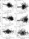
Correlations of free thyroxine (T4) and log-transformed thyrotropin (TSH) with cortisol, C-reactive protein (CRP), and albumin in coronavirus disease 2019 (COVID-19)–positive patients. Scatterplots of A, C, and E, log-transformed TSH, and B, D, and F, free T4 on the y axes are plotted against A and B, log2 cortisol, C and D, log2 CRP, and E and F, albumin on the x axes. Pearson correlation is shown at the top with its associated P value. Regression line (black line) and 95% CI (gray shading) are plotted.
Figure 3.
Figure 3.
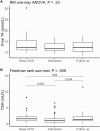
Longitudinal study of thyrotropin (TSH) and free thyroxine (T4) using 2019 baseline, admission, and follow up measurements. A (n = 20), free T4, and B (n = 50), TSH measurements, are plotted against the time point at which they were taken (baseline in 2019, admission with coronavirus disease 2019 (COVID-19), and follow-up). Repeated-measures one-way analysis of variance or Friedman rank sum test performed as indicated. Pairwise comparisons of TSH were performed with paired Wilcoxon signed-rank tests, with Bonferroni adjustment.

References

    1. Wei L, Sun S, Xu CH, et al. . Pathology of the thyroid in severe acute respiratory syndrome. Hum Pathol. 2007;38(1): 95-102. - PMC - PubMed
    1. Ding Y, He L, Zhang Q, et al. . Organ distribution of severe acute respiratory syndrome (SARS) associated coronavirus (SARS-CoV) in SARS patients: implications for pathogenesis virus transmission pathways. J Pathol. 2004;203(2): 622-630. - PMC - PubMed
    1. Wei L, Sun S, Zhang J, et al. . Endocrine cells of the adenohypophysis in severe acute respiratory syndrome (SARS). Biochem Cell Biol. 2010;88(4):723-730. - PubMed
    1. Li H, Liu SM, Yu XH, Tang SL, Tang CK. Coronavirus disease 2019 (COVID-19): current status and future perspectives. Int J Antimicrob Agents. 2020;55(5):105951. - PMC - PubMed
    1. Brancatella A, Ricci D, Cappellani D, et al. . Is subacute thyroiditis an underestimated manifestation of SARS-CoV-2 infection? Insights from a case series. [Published online ahead of print August 11, 2020.] J Clin Endocrinol Metab. doi: 10.1210/clinem/dgaa537 - DOI - PMC - PubMed

Publication types

MeSH terms