

Intraspinal stimulation with a silicon-based 3D chronic microelectrode array for bladder voiding in cats
- PMID: 33181490
- PMCID: PMC8113353
- DOI: 10.1088/1741-2552/abca13
Intraspinal stimulation with a silicon-based 3D chronic microelectrode array for bladder voiding in cats
Abstract
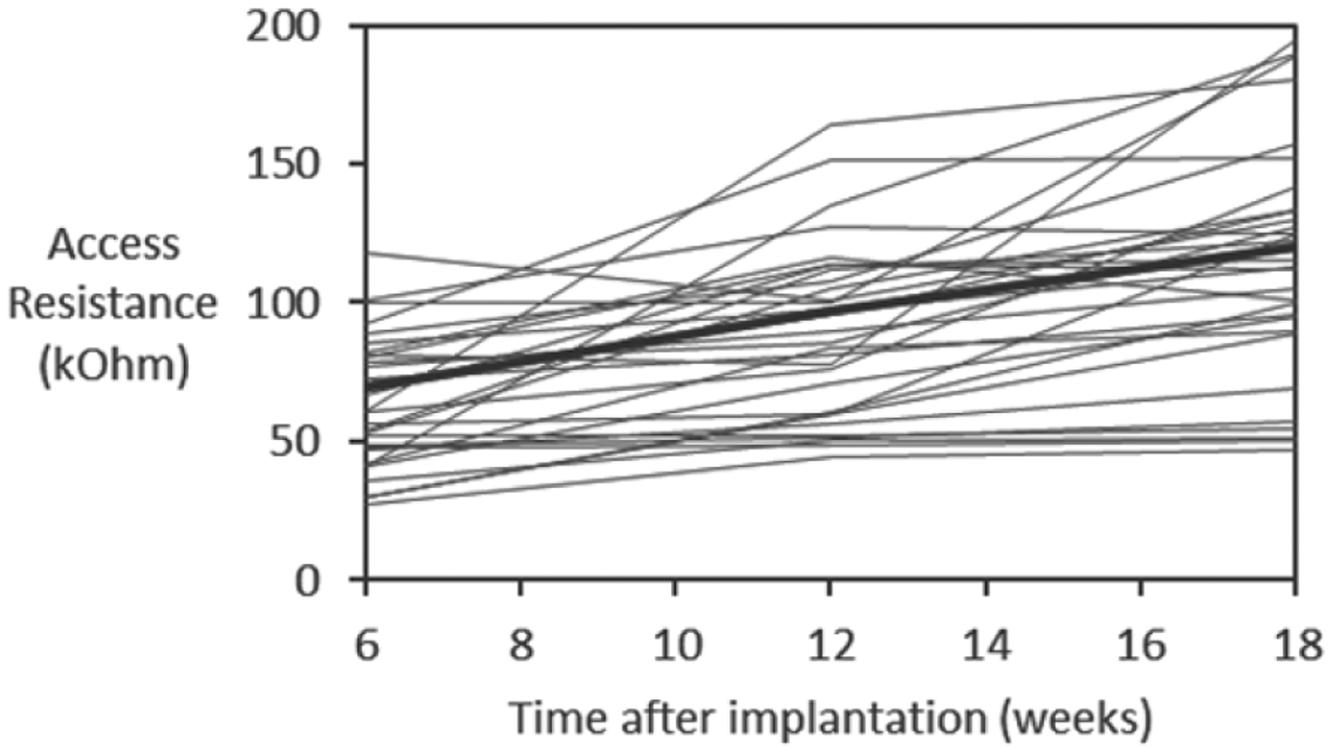
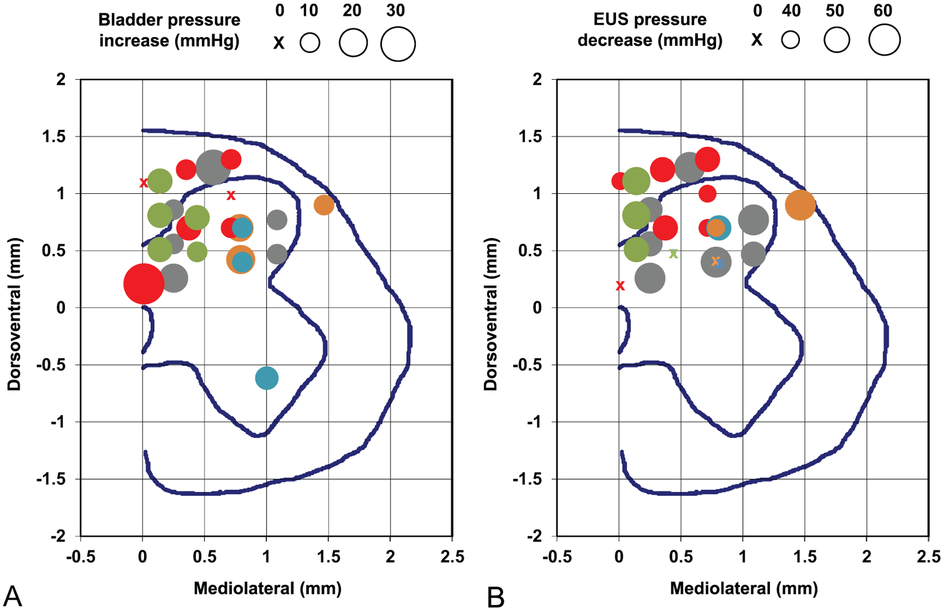
Objective.Bladder dysfunction is a significant and largely unaddressed problem for people living with spinal cord injury (SCI). Intermittent catheterization does not provide volitional control of micturition and has numerous side effects. Targeted electrical microstimulation of the spinal cord has been previously explored for restoring such volitional control in the animal model of experimental SCI. Here, we continue the development of the intraspinal microstimulation array technology to evaluate its ability to provide more focused and reliable bladder control in the feline animal model.Approach.For the first time, a mechanically robust intraspinal multisite silicon array was built using novel microfabrication processes to provide custom-designed tip geometry and 3D electrode distribution. Long-term implantation was performed in eight spinally intact animals for a period up to 6 months, targeting the dorsal gray commissure area in the S2 sacral cord that is known to be involved in the coordination between the bladder detrusor and the external urethral sphincter.Main results.About one third of the electrode sites in the that area produced micturition-related responses. The effectiveness of stimulation was further evaluated in one of eight animals after spinal cord transection (SCT). We observed increased bladder responsiveness to stimulation starting at 1 month post-transection, possibly due to supraspinal disinhibition of the spinal circuitry and/or hypertrophy and hyperexcitability of the spinal bladder afferents.Significance. 3D intraspinal microstimulation arrays can be chronically implanted and provide a beneficial effect on the bladder voiding in the intact spinal cord and after SCT. However, further studies are required to assess longer-term reliability and safety of the developed intraspinal microstimulation array prior to eventual human translation.
Keywords: bladder dysfunction; microstimulation; silicon-based microelectrode array; spinal cord injury.
Creative Commons Attribution license.
Figures


Similar articles
-
Bladder and urethral sphincter responses evoked by microstimulation of S2 sacral spinal cord in spinal cord intact and chronic spinal cord injured cats.Exp Neurol. 2004 Nov;190(1):171-83. doi: 10.1016/j.expneurol.2004.07.001. Exp Neurol. 2004. PMID: 15473990
-
Intraspinal stimulation for bladder voiding in cats before and after chronic spinal cord injury.J Neural Eng. 2007 Dec;4(4):356-68. doi: 10.1088/1741-2560/4/4/002. Epub 2007 Oct 2. J Neural Eng. 2007. PMID: 18057503 Free PMC article.
-
Arrays for chronic functional microstimulation of the lumbosacral spinal cord.IEEE Trans Neural Syst Rehabil Eng. 2004 Jun;12(2):195-207. doi: 10.1109/TNSRE.2004.827223. IEEE Trans Neural Syst Rehabil Eng. 2004. PMID: 15218934
-
Control of urinary bladder function with devices: successes and failures.Prog Brain Res. 2006;152:163-94. doi: 10.1016/S0079-6123(05)52011-9. Prog Brain Res. 2006. PMID: 16198700 Review.
-
Neurochemical plasticity and the role of neurotrophic factors in bladder reflex pathways after spinal cord injury.Prog Brain Res. 2006;152:97-115. doi: 10.1016/S0079-6123(05)52007-7. Prog Brain Res. 2006. PMID: 16198696 Review.
Cited by
-
Charge Injection Enhancement Comparisons of Iridium Oxide Microelectrodes In Vitro and In Vivo Using a Portable Neurostimulator.Int IEEE EMBS Conf Neural Eng. 2023 Apr;2023:10.1109/ner52421.2023.10123832. doi: 10.1109/ner52421.2023.10123832. Epub 2023 May 19. Int IEEE EMBS Conf Neural Eng. 2023. PMID: 38590827 Free PMC article.
-
Maximizing Charge Injection Limits of Iridium Oxide Electrodes with a Programmable Anodic Bias Circuit.Int IEEE EMBS Conf Neural Eng. 2021 May;2021:540-543. doi: 10.1109/ner49283.2021.9441282. Epub 2021 Jun 2. Int IEEE EMBS Conf Neural Eng. 2021. PMID: 34925702 Free PMC article.
-
A Diagnostic Circuit for Crosstalk Detection in Microelectrode Arrays.Int IEEE EMBS Conf Neural Eng. 2021 May;2021:544-547. doi: 10.1109/ner49283.2021.9441164. Epub 2021 Jun 2. Int IEEE EMBS Conf Neural Eng. 2021. PMID: 34925703 Free PMC article.
-
Recent progress and challenges in the treatment of spinal cord injury.Protein Cell. 2023 Sep 14;14(9):635-652. doi: 10.1093/procel/pwad003. Protein Cell. 2023. PMID: 36856750 Free PMC article. Review.
-
The inhibitory effect of intraspinal microstimulation of the sacral spinal cord on nonlinear bladder reflex dynamics in cats.Front Neurosci. 2025 Feb 3;19:1519377. doi: 10.3389/fnins.2025.1519377. eCollection 2025. Front Neurosci. 2025. PMID: 39963259 Free PMC article.
References
-
- National Spinal Cord Injury Statistical Center. Spinal cord injury: facts and figures at a glance. 2020.
-
- Anderson KD 2004. Targeting recovery: priorities of the spinal cord-injured population J. Neurotrauma 21 1371–83 - PubMed
-
- Anderson CE, Chamberlain JD, Jordan X, Kessler TM, Luca E, Möhr S, Pannek J, Schubert M, Brinkhof MWG and Swi SCISG 2019. Bladder emptying method is the primary determinant of urinary tract infections in patients with spinal cord injury: results from a prospective rehabilitation cohort study BJU Int. 123 342–52 - PubMed
-
- Roth JD, Pariser JJ, Stoffel JT, Lenherr SM, Myers JB, Welk B and Elliott SP 2019. Patient subjective assessment of urinary tract infection frequency and severity is associated with bladder management method in spinal cord injury Spinal Cord 57 700–7 - PubMed
-
- Tofte N, Nielsen ACY, Trøstrup H, Andersen CB, Von Linstow M, Hansen B, Biering-Sørensen F, Høiby N and Moser C 2017. Chronic urinary tract infections in patients with spinal cord lesions—biofilm infection with need for long-term antibiotic treatment APMIS 125 385–91 - PubMed
Publication types
MeSH terms
Substances
Grants and funding
LinkOut - more resources
Full Text Sources
Medical
Miscellaneous