. 2020 Nov 9;8(11):484.
doi: 10.3390/biomedicines8110484.

Induction of Durable Antitumor Response by a Novel Oncolytic Herpesvirus Expressing Multiple Immunomodulatory Transgenes

Affiliations

Induction of Durable Antitumor Response by a Novel Oncolytic Herpesvirus Expressing Multiple Immunomodulatory Transgenes

Dmitry V Chouljenko et al. Biomedicines. .

Abstract

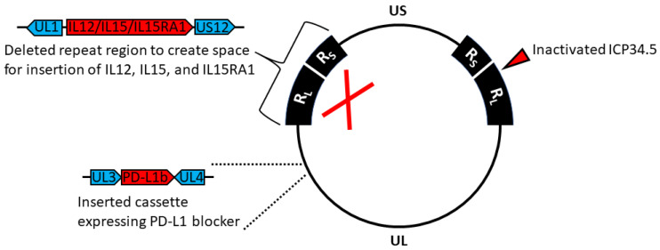
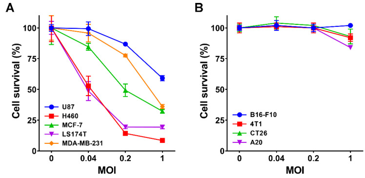
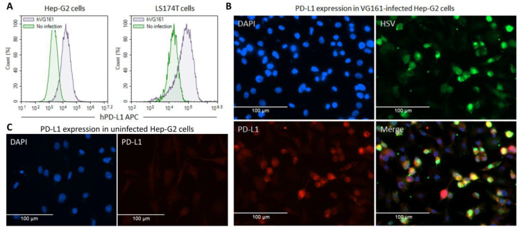
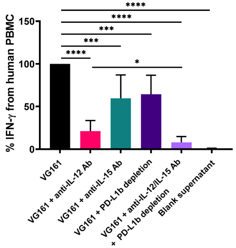
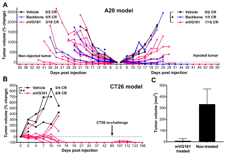
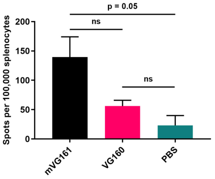
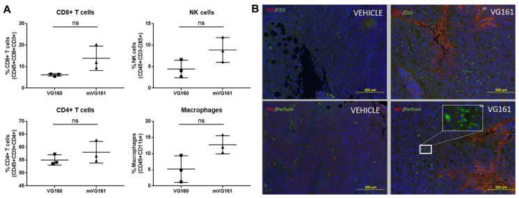
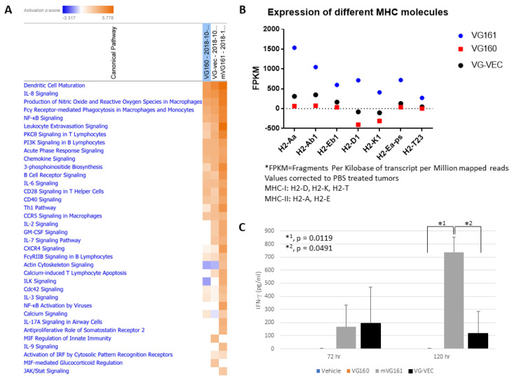
Oncolytic virotherapy is a promising new tool for cancer treatment, but direct lytic destruction of tumor cells is not sufficient and must be accompanied by strong immune activation to elicit anti-tumor immunity. We report here the creation of a novel replication-competent recombinant oncolytic herpes simplex virus type 1 (VG161) that carries genes coding for IL-12, IL-15, and IL-15 receptor alpha subunit, along with a peptide fusion protein capable of disrupting PD-1/PD-L1 interactions. The VG161 virus replicates efficiently and exhibits robust cytotoxicity in multiple tumor cell lines. Moreover, the encoded cytokines and the PD-L1 blocking peptide work cooperatively to boost immune cell function. In vivo testing in syngeneic CT26 and A20 tumor models reveals superior efficacy when compared to a backbone virus that does not express exogenous genes. Intratumoral injection of VG161 induces abscopal responses in non-injected distal tumors and grants resistance to tumor re-challenge. The robust anti-tumor effect of VG161 is associated with T cell and NK cell tumor infiltration, expression of Th1 associated genes in the injection site, and increased frequency of splenic tumor-specific T cells. VG161 also displayed a superb safety profile in GLP acute and repeated injection toxicity studies performed using cynomolgus monkeys. Overall, we demonstrate that VG161 can induce robust oncolysis and stimulate a robust anti-tumor immune response without sacrificing safety.

Keywords: VG161; antitumor immunity; cancer vaccine; combinatorial therapy; herpes simplex virus; immune checkpoint blockade; immunotherapy; interleukin-12; interleukin-15; oncolytic virus.


Conflict of interest statement

All authors are employed by, and have ownership interest (including stock, patents, etc.) in Virogin Biotech Canada Ltd. Patent applications have been filed to cover VG161 and related technologies. This study was wholly funded by Virogin Biotech Canada Ltd., but the study authors retained absolute discretion over the design and execution of the study, the collection, analysis and interpretation of the data, and the writing of the manuscript. Study funding was not conditioned on the outcome of the research.

Figures

Figure 1
Figure 1
Genomic map of VG161. Prototypic arrangement of the wild-type HSV-1 genome with the unique long (UL) and unique short (US) regions flanked by inverted repeats RL and RS, respectively. Expanded regions indicate modifications made to the HSV-1 genome during construction of VG161.
Figure 2
Figure 2
Cytotoxicity of mutant viruses. (A) The cytotoxic effect of VG161 virus was evaluated in a variety of human cancer cell monolayers including U87, H460, MCF-7, LS174T, and MDA-MB-231 at 72 h post infection and MOI of 0.04, 0.2, and 1. Cell survival percentage was quantified by MTT assay. (B) A panel of 4 different mouse tumor cell lines including B16-F10, 4T1, CT26, and A20 was infected with mVG161 virus at MOI 0, 0.04, 0.2, and 1. Cell viability was quantified using MTT assay at 72 h post infection. Error bars indicate SD.
Figure 3
Figure 3
Infection with VG161 leads to upregulation of PD-L1 expression. (A) Hep-G2 and LS174T cells were seeded in a 12-well plate (2 × 10⁵ cells/well) and cultured at 5% CO2 and 37 °C overnight. The next day, half of the seeded cells were infected with hVG161 virus (MOI = 1) for 24 h. Cells were subsequently harvested and immunostained with purified rabbit monoclonal anti-human PD-L1 antibody plus APC-conjugated anti-rabbit IgG antibody and the expression level of PD-L1 was assessed by flow cytometry. (B,C) 5 × 10⁴/well of Hep-G2 cells were seeded in a 24 well plate with coverslip and incubated overnight at 37°C, followed by infection with hVG161 at MOI = 1 for 6 h. Cells were fixed in 4% PFA for 5 min and incubated in 3% skim milk in PBS-T for 30 min, followed by an overnight incubation at 4 °C with monoclonal mouse anti-HSV antibody (Abcam, 1:100 dilution) and with polyclonal rabbit anti-PDL1 antibody (Abcam, 1:100 dilution). The fixed cells were subsequently incubated with a mixture of fluorophore-labeled secondary antibodies (Alexa Fluor 488 goat anti-mouse and Alexa Fluor 568 goat anti-rabbit, Invitrogen, Burlington, ON, Canada; 1:500 dilution) in the dark for 1 h, counterstained with Hoechst (Sigma), and mounted on glass slides for imaging. (B) VG161-infected Hep-G2 cells. (C) Uninfected Hep-G2 cells.
Figure 4
Figure 4
IL-12, IL-15/IL-15RA, and PD-L1 blocker cooperatively enhance immune cell function in vitro. PHA-activated human PBMCs were co-incubated with recombinant human PD-L1 protein and supernatant from VG161-infected Vero cells for 48 h. Antibody-mediated neutralization of IL-12 and/or IL-15 was carried out in conjunction with depletion of PD-L1 blocker. Co-incubation with supernatant from uninfected cells was used as negative control (blank supernatant). Human IFN-γ production was assessed by ELISA. * p < 0.05, *** p < 0.001, **** p < 0.0001. p values were computed using unpaired t-test. Error bars indicate SD.
Figure 5
Figure 5
In vivo efficacy of VG161 following intratumoral inoculation. Three nude mice per group were subcutaneously implanted with 2 × 10⁶ LS174T human colon adenocarcinoma cells into the lower right flank, followed by a single intratumoral injection of either vehicle (PBS) control or 5 × 10⁵ or 5 × 10⁶ PFU/mouse of VG161. * p < 0.05, ** p <0.01. p values were computed using unpaired t-test.
Figure 6
Figure 6
Virus biodistribution. Nude mice bearing LS174T tumors were injected intratumorally with either 1 or 3 doses of VG161 (1 dose = 5 × 10⁷ PFU/mouse). Mice were euthanized at different time points, and genomic DNA was isolated from these organs and subjected to qPCR to quantify the viral copy number using the codon optimized IL-15RA1 gene due to its specificity to VG161.
Figure 7
Figure 7
In vivo efficacy of a murine version of VG161 (mVG161) following intratumoral inoculation. (A) A20 cells were subcutaneously implanted into immunocompetent BALB/C mice in both sides of lower flanks (5 × 10⁶ A20 cells per flank). 5 × 10⁶ PFU/mouse/day of either mVG161 or VG160 backbone virus (version of VG161 without payload) was injected once per day for 5 consecutive days into tumors on one side only. 16 mice were treated with mVG161, 5 mice were treated with VG160, and 2 mice were treated with vehicle (PBS) control. (B) Thirteen immunocompetent BALB/C mice were subcutaneously implanted with 1 × 10⁶ CT26 cells/mouse into the lower flanks, with 9 animals randomly assigned to the mVG161 treatment group (5 × 10⁶ PFU/mouse injected 5 times) and another 4 animals to the vehicle (PBS) control group. At 90 days post injection, the surviving 6 mice in the mVG161-treated group were re-implanted with 1 × 10⁶ CT26 cells in the same location. (C) Tumor sizes 7 days after CT26 re-challenge in mVG161-treated animals compared to age-matched (28 weeks old) control mice that were not treated with mVG161. CR = complete response. * p < 0.05, ** p < 0.01. p values were computed using unpaired t-test. Error bars indicate SD.
Figure 8
Figure 8
ELISpot assay to evaluate T cell activity in spleens from tumor bearing mice treated with mVG161. BALB/c mice were subcutaneously implanted with 1 × 10⁶ CT26 tumor cells into the lower right flank, followed by multiple injections of 5 × 10⁶ PFU/mouse/day, for 5 consecutive days, of either mVG161, VG160, or PBS control. Mouse IFN-γ ELISpot assay was performed on splenocytes collected from CT26 tumor bearing mice at 5, 7, and 9 days post-injection and exposed to CT26 cells (results shown are from 5 days post treatment). Quantitative results are graphed with 2 mice per group. p values were computed using unpaired t-test. Error bars indicate SD.
Figure 9
Figure 9
Effect of mVG161 treatment on intratumoral lymphocyte populations. BALB/c mice were subcutaneously implanted with 1 × 10⁶ CT26 tumor cells, followed 8 days later by 5 consecutive injections of PBS (vehicle), VG160 backbone, or mVG161 virus (5 × 10⁶ PFU/mouse/day). Tumors were harvested 24 h after final injection. (A) Percentages of different subsets of T cells, NK cells, and macrophages within the tumor mass were analyzed by flow cytometry by gating on CD45+ leukocytes and then looking at the population of CD8+/CD4+ T cells, NK cells, and macrophages based on different surface markers (ns = not significant). (B) Immunohistochemical analysis was performed on sections of excised CT26 tumor treated with either PBS control (vehicle) or with mVG161 using monoclonal antibodies against CD3 and perforin and polyclonal antibodies against HSV-1. p values were computed using unpaired t-test.
Figure 10
Figure 10
VG161 multi-factor payload elicits a stronger immune response than GM-CSF. (A) BALB/c mice were subcutaneously implanted with 1 × 10⁶ CT26 tumor cells and subsequently treated with 5 daily injections (5 × 10⁶ PFU/mouse/day) of VG161, VG160 (backbone) or VG-VEC (VG160 expressing GM-CSF). Tumors were harvested 24 h after the final virus injection, RNA was isolated and purified, followed by transcriptome sequencing using the Illumina NGS platform. Data were analyzed using Qiagen Ingenuity Pathway Analysis (IPA) software to evaluate activation of immunostimulatory pathways in each treatment group. (B) Expression of MHC molecules in each treatment group was also quantified, and the over-expression of some MHC targets was validated by RT-qPCR. (C) BALB/c mice were implanted with 1 × 10⁶ CT26 tumor cells and injected 5 times daily with the indicated viruses (5 × 10⁶ PFU/mouse/day). Mouse serum was collected either 72 h or 120 h after the final injection, and mouse IFN-γ production was assessed by ELISA assay. p values were computed using an unpaired t-test. Error bars indicate SD.

References

    1. Kelly E., Russell S.J. History of oncolytic viruses: Genesis to genetic engineering. Mol. Ther. 2007;15:651–659. doi: 10.1038/sj.mt.6300108. - DOI - PubMed
    1. Lawler S.E., Speranza M.-C., Cho C.-F., Chiocca E.A. Oncolytic Viruses in Cancer Treatment: A Review. JAMA Oncol. 2017;3:841–849. doi: 10.1001/jamaoncol.2016.2064. - DOI - PubMed
    1. Liu B.L., Robinson M., Han Z.-Q., Branston R.H., English C., Reay P., McGrath Y., Thomas S.K., Thornton M., Bullock P., et al. ICP34.5 deleted herpes simplex virus with enhanced oncolytic, immune stimulating, and anti-tumour properties. Gene Ther. 2003;10:292–303. doi: 10.1038/sj.gt.3301885. - DOI - PubMed
    1. Hu J.C.C., Coffin R.S., Davis C.J., Graham N.J., Groves N., Guest P.J., Harrington K.J., James N.D., Love C.A., McNeish I., et al. A phase I study of OncoVEXGM-CSF, a second-generation oncolytic herpes simplex virus expressing granulocyte macrophage colony-stimulating factor. Clin. Cancer Res. 2006;12:6737–6747. doi: 10.1158/1078-0432.CCR-06-0759. - DOI - PubMed
    1. Andtbacka R.H.I., Kaufman H.L., Collichio F., Amatruda T., Senzer N., Chesney J., Delman K.A., Spitler L.E., Puzanov I., Agarwala S.S., et al. Talimogene Laherparepvec Improves Durable Response Rate in Patients With Advanced Melanoma. J. Clin. Oncol. 2015;33:2780–2788. doi: 10.1200/JCO.2014.58.3377. - DOI - PubMed