

Diagnostic Value of Unenhanced CT Attenuation and CT Histogram Analysis in Differential Diagnosis of Adrenal Tumors
- PMID: 33182333
- PMCID: PMC7695290
- DOI: 10.3390/medicina56110597
Diagnostic Value of Unenhanced CT Attenuation and CT Histogram Analysis in Differential Diagnosis of Adrenal Tumors
Abstract
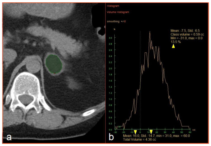
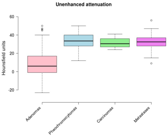
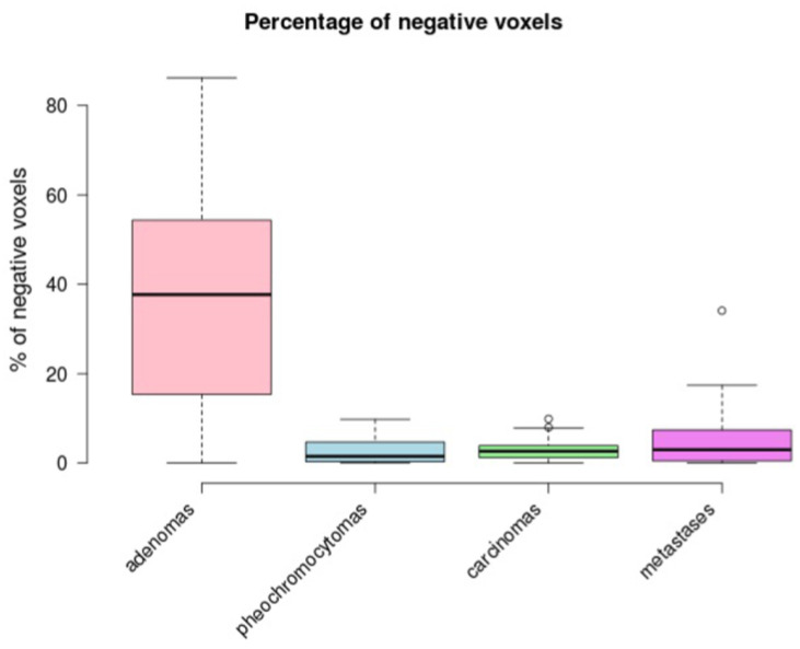
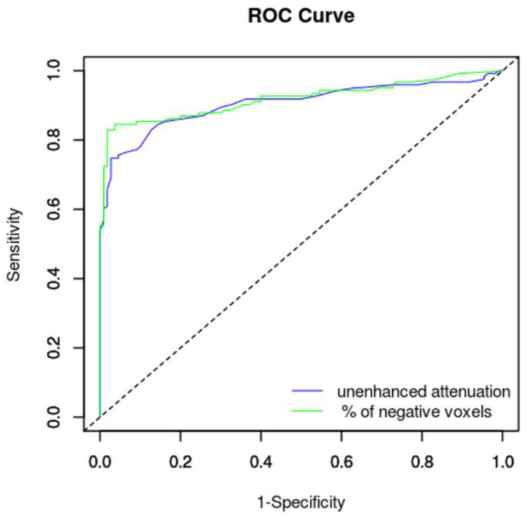
Background and Objectives: Our aim was to verify the optimal cut-off value for unenhanced CT attenuation and the percentage of negative voxels in the volume CT histogram analysis of adrenal masses. Materials and Methods: We retrospectively analyzed the CT data of patients who underwent an adrenalectomy in the period 2002-2019. In total, 413 adrenalectomies were performed. Out of these, 233 histologically verified masses (123 adenomas, 58 pheochromocytomas, 18 carcinomas, and 34 metastases) fulfilled the inclusion criteria and were selected for analysis. The mean unenhanced attenuation in Hounsfield units (HU) and the percentage of voxels with attenuation less than 0 HU (negative voxels) were measured in each mass. Results: The mean unenhanced attenuation with a cut-off value of 10 HU reached a sensitivity of 59.4% and a specificity of 99.1% for benign adenomas. The mean unenhanced attenuation with a cut-off value of 15 HU reached a sensitivity of 69.1% and a specificity of 98.2%. For the histogram analysis, a cut-off value of 10% of negative pixels reached a sensitivity of 82.9% and a specificity of 98.2%, whereas a cut-off value of 5% of negative pixels reached a sensitivity of 87.8% and a specificity of 75.5%. The percentage of negative voxels reached a slightly better area under the curve (0.919) than unenhanced attenuation (0.908). Conclusion: Mean unenhanced attenuation with a cut-off value of 10 HU represents a simple tool, and the most specific one, to distinguish adrenal adenomas from non-adenomas. CT histogram analysis with cut-off values of 10% of negative voxels improves sensitivity without any loss of specificity.
Keywords: adrenal gland neoplasms; adrenal incidentaloma; adrenocortical adenoma; computed tomography; histogram analysis.
Conflict of interest statement
The authors declare no conflict of interest.
Figures
References
-
- Čtvrtlík F., Heřman M., Študent V., Tichá V., Minařík J. Differential diagnosis of incidentally detected adrenal masses revealed on routine abdominal CT. Eur. J. Radiol. 2009;69:243–252. - PubMed
MeSH terms
Grants and funding
LinkOut - more resources
Full Text Sources
Medical
