

Clinical and Molecular-Genetic Insights into the Role of Oxidative Stress in Diabetic Retinopathy: Antioxidant Strategies and Future Avenues
- PMID: 33182408
- PMCID: PMC7697026
- DOI: 10.3390/antiox9111101
Clinical and Molecular-Genetic Insights into the Role of Oxidative Stress in Diabetic Retinopathy: Antioxidant Strategies and Future Avenues
Abstract
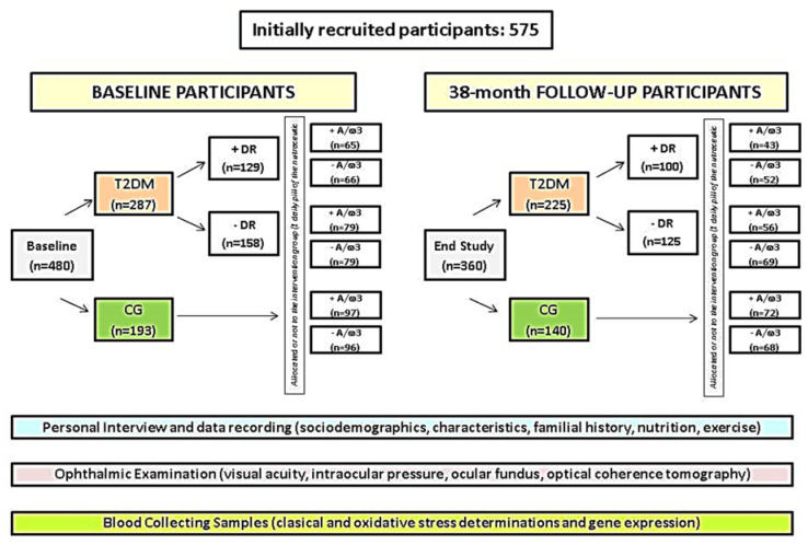
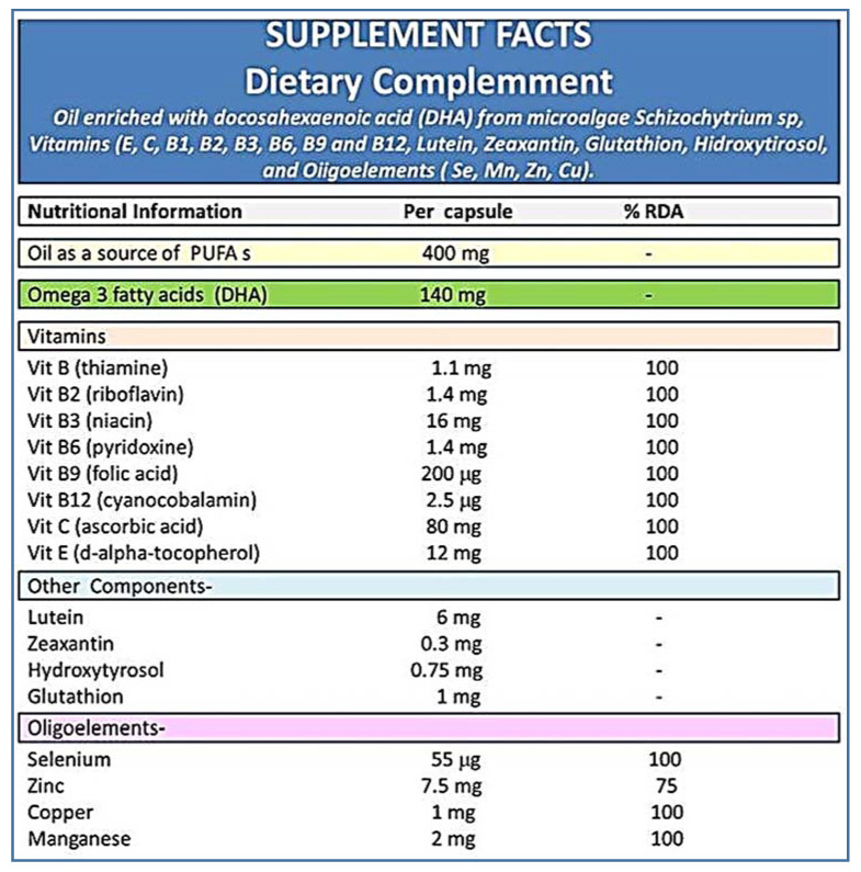
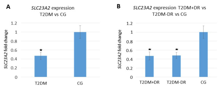
Reactive oxygen species (ROS) overproduction and ROS-signaling pathways activation attack the eyes. We evaluated the oxidative stress (OS) and the effects of a daily, core nutritional supplement regimen containing antioxidants and omega 3 fatty acids (A/ω3) in type 2 diabetics (T2DM). A case-control study was carried out in 480 participants [287 T2DM patients with (+)/without (-) diabetic retinopathy (DR) and 193 healthy controls (CG)], randomly assigned to a daily pill of A/ω3. Periodic evaluation through 38 months allowed to outline patient characteristics, DR features, and classic/OS blood parameters. Statistics were performed by the SPSS 24.0 program. Diabetics displayed significantly higher circulating pro-oxidants (p = 0.001) and lower antioxidants (p = 0.0001) than the controls. Significantly higher plasma malondialdehyde/thiobarbituric acid reactive substances (MDA/TBARS; p = 0.006) and lower plasma total antioxidant capacity (TAC; p = 0.042) and vitamin C (0.020) was found in T2DM + DR versus T2DM-DR. The differential expression profile of solute carrier family 23 member 2 (SLC23A2) gene was seen in diabetics versus the CG (p = 0.001), and in T2DM + DR versus T2DM - DR (p < 0.05). The A/ω3 regime significantly reduced the pro-oxidants (p < 0.05) and augmented the antioxidants (p < 0.05). This follow-up study supports that a regular A/ω3 supplementation reduces the oxidative load and may serve as a dietary prophylaxis/adjunctive intervention for patients at risk of diabetic blindness.
Keywords: antioxidants; candidate biomarkers; omega-3 fatty acids; oxidative stress; prevention of blindness; retinopathy; type 2 diabetes mellitus.
Conflict of interest statement
All authors of this work have disclosed that they have no significant financial relationships or financial interests in the commercial companies that are related to this study or paper.
Figures
References
-
- Chawla R., Madhu S.V., Makkar B.M., Ghosh S., Saboo B., Kalra S. On behalf of RSSDI-ESI Consensus Group RSSDI-ESI clinical practice recommendations for the management of Type 2 diabetes mellitus 2020. Int. J. Diabetes Dev. Ctries. 2020;40(Suppl. S1–S122):1–122. doi: 10.1007/s13410-020-00819-2. - DOI - PMC - PubMed
-
- Hammes H.P., Welp R., Kempe H.P., Wagner C., Siegel E., Holl R.W. DPV initiative—German BMBF competence network diabetes mellitus. Risk factors for retinopathy and diabetic macular edema in Type 2 diabetes-results from the German/Austrian DPV Database. PLoS ONE. 2015;10:e0132492. doi: 10.1371/journal.pone.0132492. - DOI - PMC - PubMed
Grants and funding
LinkOut - more resources
Full Text Sources
