. 2020 Nov 9;12(11):1281.
doi: 10.3390/v12111281.

HIV-1 Gag Forms Ribonucleoprotein Complexes with Unspliced Viral RNA at Transcription Sites

Affiliations

HIV-1 Gag Forms Ribonucleoprotein Complexes with Unspliced Viral RNA at Transcription Sites

Kevin M Tuffy et al. Viruses. .

Abstract

The ability of the retroviral Gag protein of Rous sarcoma virus (RSV) to transiently traffic through the nucleus is well-established and has been implicated in genomic RNA (gRNA) packaging Although other retroviral Gag proteins (human immunodeficiency virus type 1, HIV-1; feline immunodeficiency virus, FIV; Mason-Pfizer monkey virus, MPMV; mouse mammary tumor virus, MMTV; murine leukemia virus, MLV; and prototype foamy virus, PFV) have also been observed in the nucleus, little is known about what, if any, role nuclear trafficking plays in those viruses. In the case of HIV-1, the Gag protein interacts in nucleoli with the regulatory protein Rev, which facilitates nuclear export of gRNA. Based on the knowledge that RSV Gag forms viral ribonucleoprotein (RNPs) complexes with unspliced viral RNA (USvRNA) in the nucleus, we hypothesized that the interaction of HIV-1 Gag with Rev could be mediated through vRNA to form HIV-1 RNPs. Using inducible HIV-1 proviral constructs, we visualized HIV-1 Gag and USvRNA in discrete foci in the nuclei of HeLa cells by confocal microscopy. Two-dimensional co-localization and RNA-immunoprecipitation of fractionated cells revealed that interaction of nuclear HIV-1 Gag with USvRNA was specific. Interestingly, treatment of cells with transcription inhibitors reduced the number of HIV-1 Gag and USvRNA nuclear foci, yet resulted in an increase in the degree of Gag co-localization with USvRNA, suggesting that Gag accumulates on newly synthesized viral transcripts. Three-dimensional imaging analysis revealed that HIV-1 Gag localized to the perichromatin space and associated with USvRNA and Rev in a tripartite RNP complex. To examine a more biologically relevant cell, latently infected CD4+ T cells were treated with prostratin to stimulate NF-κB mediated transcription, demonstrating striking localization of full-length Gag at HIV-1 transcriptional burst site, which was labelled with USvRNA-specific riboprobes. In addition, smaller HIV-1 RNPs were observed in the nuclei of these cells. These data suggest that HIV-1 Gag binds to unspliced viral transcripts produced at the proviral integration site, forming vRNPs in the nucleus.

Keywords: HIV-1 Gag; Rev; nucleus; viral RNA.


Conflict of interest statement

The authors declare no conflict of interest.

Figures

Figure 1
Figure 1
HIV-1 Gag foci are present in the nucleus of HeLa and U2OS cells regardless of the nature of the tag fused to the C-terminus of Gag, which was expressed in a Rev-dependent fashion using the constructs shown in panels A and B. (A) Schematic of doxycycline-inducible HIV.Gag-GFP rtTA proviral construct expressing Gag fused to GFP, which is stably integrated in HeLa cells (HeLa HIV.Gag-GFP rtTA). The 5′LTR contains a mutated TAR, two NF-κB sites, eight copies of the tetO sites in the promoter, and three SP1 sites. (B) Schematic of pHIV.Gag-CFP/SNAP-tag rtTA doxycycline-inducible construct with the same 5′UTR sequences described in panel A, which expresses Gag fused to either CFP or SNAP-tag. (C) HeLa HIV.Gag-GFP rtTA cell line contains the stably-integrated proviral construct shown in panel A. Numerous Gag foci (green) were seen in the nucleus, which was stained with DAPI (blue). Three-dimensional reconstruction of a confocal z-series is shown with crosshairs through a single Gag focus to demonstrate that Gag was present inside the nucleus (outlined by a dashed white line) in all three planes. (D) HeLa cells co-expressing pHIV.Gag-CFP rtTA with pPB-t-rtTA. (E) U2OS cells transfected with pHIV.Gag-CFP rtTA and pPB-t-rtTA. (F) A HeLa cell line stably expressing rtTA was transfected with pHIV.Gag-SNAP-tag rtTA and incubated with SNAP-tag ligand JF549.
Figure 2
Figure 2
Presence of HIV-1 Gag in cytoplasmic and nuclear subcellular fractions. (A) Hela cells transfected with CMV HIV-1 Gag-GFP and EGFP-C1 plasmids were fractionated into cytoplasmic (Cyto), nucleoplasm (Nuc), and nuclear pellet (Nuc pellet) and analyzed by immunoblot using anti-p24 and anti-GFP antibodies. EGFP-C1 serves as negative control. Fraction purity was assessed using Calnexin (cytoplasm), MED4 (nucleoplasm), and fibrillarin (nucleolar, nuclear pellet). (B) Immunoblot of HeLa rtTA cell cytoplasmic and nuclear fractions following transfection with either pHIV.Gag-CFP rtTA, pHIV.Gag-SNAP-tag rtTA, or ECFP-N1 compared to untransfected cells. Fractions were probed using anti-p24 and anti-GFP antibodies for the presence of HIV-1 Gag. ECFP-N1 transfection and untransfected samples serves a negative control. Purity of the fractions was assessed using Calnexin (cytoplasmic) and MED4 (nuclear) antibodies.
Figure 3
Figure 3
HIV-1 Gag co-localizes with USvRNA in the nuclei of HeLa HIV.Gag-GFP rtTA cells. (A) Two different cells (panels i and ii) showing co-localization of HIV-1 Gag-GFP (green), USvRNA (red, detected by smFISH), and co-localized foci (yellow) in the nucleus (DAPI-stained, shown as blue, outlined in white), cytoplasm, and along the plasma membrane of dox-induced HIV-1 Gag-GFP rtTA cells. (B) Cross-sections of dox-induced HeLa HIV.Gag-GFP rtTA cells containing nuclear HIV-1 Gag-GFP foci co-localized with USvRNA within intranuclear and extranuclear space, as indicated. Gag-USVRNA co-localized foci located at the intersection of the white cross hairs) are depicted in X, Y (center); Y, Z (right); and X, Z (bottom) planes, with the DAPI-stained nucleus (blue) outlined with a white dashed line. (C) Left, Three-dimensional surface rendering exhibiting HIV-1 Gag (green) and USvRNA (red) co-localization within a single orthogonal clipping through the XY plane of the nucleus. Right, Co-localization analysis of Gag-USvRNA co-localized foci (white) using Imaris. Images were depicted in the XY (center) and YZ (rotated 90°) planes with scale bars = 2 µm (white). vRNP foci can be seen located in the perichromatin (DAPI-poor) space. (D) GAPDH RNA (detected using smFISH; red) and HIV-1 Gag-GFP (green) have negligible co-localization within the nucleus (DAPI-blue, white outline).
Figure 4
Figure 4
HIV-1 Gag specifically associates with USvRNA in nuclear and cytoplasmic fractions. (A) HeLa HIV.Gag-GFP rtTA cell fractions were immunoprecipitated using anti-GFP antibodies and detected for the presence of HIV-1 Gag via p24. Anti-IgG immunoprecipitation serves as negative control. Fraction purity was assessed using Lamin A/C and α-tubulin. (B) Immunoblot analysis of HIV-1 Gag-GFP immunoprecipitated from cytoplasmic and nuclear fractions of induced HeLa HIV.Gag-GFP rtTA cells. RT-qPCR analysis of targeted unspliced vRNAs (US), singly-spliced vRNAs (SS), multiply-spliced vRNAs (MS), and beta-actin RNAs with HIV-1 Gag-GFP immunoprecipitated from fractionated HIV-1 Gag-GFP rtTA cells.
Figure 5
Figure 5
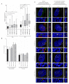
Transcription inhibition of HeLa HIV.Gag-GFP rtTA cells decreased the number of nuclear HIV-1 Gag and USvRNA foci but increased the degree of co-localization. Quantitative analysis of dox-induced HeLa HIV.Gag-GFP rtTA cells demonstrating the mean percent of nuclear HIV-1 Gag co-localization with USvRNA and USvRNA co-localization with HIV-1 Gag following transcription inhibition with DRB (A) or Act D (B) for 30 and 60 min compared to the untreated control. (error bars = standard error of the mean, statistical significances: * P < 0.05, ** P < 0.01, *** P < 0.001, **** P < 0.0001). (C) 3D surface renderings of dox-induced HeLa HIV.Gag-GFP rtTA cells depicting the co-localization (shown in white) of HIV-1 Gag-GFP (green) and USvRNA (red, detected by smFISH) foci within the nucleus (DAPI, blue) following treatment with the transcription inhibitor DRB or Act D for 30 and 60 min. 3D surface renderings were subjected to an orthogonal clipping plane to bisect the nucleus in XY or YZ planes. The white rectangle represents the region of interest enlarged below.
Figure 6
Figure 6
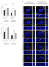
Inhibiting CRM1-mediated nuclear export in HeLa HIV.Gag-GFP rtTA cells increased HIV-1 Gag and USvRNA nuclear foci but did not affect the degree of co-localization between HIV-1 Gag and USvRNA. (A) Quantitative analysis depicting the mean number of HIV-1 Gag-GFP and USvRNA nuclear foci in dox-induced HeLa HIV.Gag-GFP rtTA cells following LMB treatment compared to the untreated control cells. (B) Quantitative analysis of dox-induced HIV-1 Gag-GFP rtTA cells demonstrating the mean percent of nuclear HIV-1 Gag co-localization with USvRNA and USvRNA co-localization with HIV-1 Gag following treatment with LMB for 30, 60, 90, or 120 min prior to fixation compared to the untreated control. (error bars = standard error of the mean, statistical significances: * P < 0.05, ** P < 0.01, *** P < 0.001, **** P < 0.0001). (C) 3D surface renderings of dox-induced HeLa HIV.Gag-GFP rtTA cells depicting the co-localization (shown in white) of HIV-1 Gag-GFP (green) and USvRNA (red, detected by FISH) foci within the nucleus (DAPI, blue) following treatment with LMB. 3D surface renderings were subjected to an orthogonal clipping plane bisected in the X, Y and Y, Z planes to show the co-localization of foci within the nucleus. The white rectangle represents the region of interest enlarged in the panels below.
Figure 7
Figure 7
HIV-1 Gag and Rev are both present in chromatin-associated fractions and are co-localized in three dimensions. (A) 293T cells transfected with HIV-1 Gag-GFP with or without Rev-YFP expression plasmids were separated into chromatin-associated protein fractions representing euchromatin (Chr 150) and heterochromatin (Chr 600) and analyzed by immunoblotting using anti-GFP antibodies. Fraction purity was assessed by immunoblotting with antibodies to detect GAPDH (present only in whole cell lysate, WCL), CRM1 (WCL and chromatin), RCC1 (WCL and chromatin) and Histone H2B (WCL and chromatin). (B) Localization of HIV-1 Gag-GFP (green) and USvRNA (red, detected by smFISH) in two dividing cells (i and ii) showing persistence of Gag-USvRNA co-localization during metaphase but no binding to the chromosome, which was stained blue using DAPI. (C) Schematic diagram of dox-inducible Rev-BFP used to create HeLa HIV.Gag-GFP rtTA Rev-BFP cells. (D) 3D-surface renderings of induced HeLa HIV.Gag-GFP rtTA Rev-BFP cells depicting three-way co-localization in merged images of Gag-GFP (green) and USvRNA (red, detected by smFISH) within the Rev-BFP (blue) mask using orthogonal clipping planes through the nucleus of 0° and 90° rotation. The large white square is the frame that denotes the cell as three-dimensional, the yellow square indicates that the cell was subjected to the orthogonal clipping plane, and the small white rectangle represents the region of interest that was enlarged in the panel below. A co-localization algorithm was used to demonstrate Gag and USvRNA co-localization (white foci) within the confines of the Rev (blue) signal. The small rectangle was enlarged below to provide additional detail.
Figure 8
Figure 8
HIV-1 Gag and co-localizes with USvRNA in the nucleus of JLat 10.6 cells. (A,B) Confocal images showing (i) single z-slices and (ii) cross-sections of three-dimensional reconstructions of two examples of JLat 10.6 cells activated by prostratin. USvRNA labeled via smFISH (red) is co-localized with full-length HIV-1 Gag (anti-p24 cy5 labeled; green) in the nucleus outlined in white dashed line (DAPI, blue). A co-localization channel (white) was generated to show that the Gag and USvRNA signals are present in the same pixels of the image. In each cell, a vRNP consisting of USvRNA and Gag was present within a crosshair to show its location within the nucleus in three planes. (C) Immunoblot of JLat 10.6 whole cell lysates in the absence (-) or presence (+) of prostratin. The full-length Gag polyprotein was present in cells reactivated from latency. GAPDH served as a loading control.
Figure 9
Figure 9
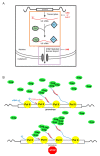
Proposed model of HIV-1 Gag association with USvRNA during transcription. (A) Our results demonstrate that treatment of HeLa HIV.Gag-GFP rtTA cells with Act D or DRB caused Gag to accumulate on USvRNAs that may be at or near transcription sites (orange box). Because Rev also binds USvRNA co-transcriptionally, there is a possibility that both Gag and Rev associate with USvRNA at transcription sites. Nuclear export of USvRNA was inhibited by LMB (purple box), along with small increase in HIV-1 Gag-GFP nuclear foci, yet there was no change in the degree of Gag-USvRNA co-localization. This finding suggests that there is a narrow “window of opportunity” for Gag-USvRNA to form vRNP complexes, starting at transcription but ending prior to formation of the export complex. (B) The model proposes that Gag binds to the 5′UTR psi sequence as it emerges during transcription. With transcription inhibition, Gag accumulates on stalled transcription complexes leading to an increase in Gag-USvRNA co-localization, potentially through additional Gag-Gag or Gag-RNA interactions.

Similar articles

Cited by

References

    1. Rabson A.B., Graves B.J. Synthesis and Processing of Viral RNA. In: Coffin J.M., Hughes S.H., Varmus H.E., editors. Retroviruses. Cold Spring Harbor Laboratory Press; Cold Spring Harbor, NY, USA: 1997. - PubMed
    1. D’Souza V., Summers M.F. How retroviruses select their genomes. Nat. Rev. Microbiol. 2005;3:643–655. doi: 10.1038/nrmicro1210. - DOI - PubMed
    1. Jouvenet N., Laine S., Pessel-Vivares L., Mougel M. Cell biology of retroviral RNA packaging. RNA Biol. 2011;8:572–580. doi: 10.4161/rna.8.4.16030. - DOI - PMC - PubMed
    1. Jouvenet N., Neil S.J., Bess C., Johnson M.C., Virgen C.A., Simon S.M., Bieniasz P.D. Plasma membrane is the site of productive HIV-1 particle assembly. PLoS Biol. 2006;4:e435. doi: 10.1371/journal.pbio.0040435. - DOI - PMC - PubMed
    1. Chen J., Rahman S.A., Nikolaitchik O.A., Grunwald D., Sardo L., Burdick R.C., Plisov S., Liang E., Tai S., Pathak V.K., et al. HIV-1 RNA genome dimerizes on the plasma membrane in the presence of Gag protein. Proc. Natl. Acad. Sci. USA. 2016;113:E201–E208. doi: 10.1073/pnas.1518572113. - DOI - PMC - PubMed

Publication types

MeSH terms

Substances