. 2020 Nov 10;12(11):3313.
doi: 10.3390/cancers12113313.

Novel FGFR4-Targeting Single-Domain Antibodies for Multiple Targeted Therapies against Rhabdomyosarcoma

Affiliations

Novel FGFR4-Targeting Single-Domain Antibodies for Multiple Targeted Therapies against Rhabdomyosarcoma

Nagjie Alijaj et al. Cancers (Basel). .

Abstract

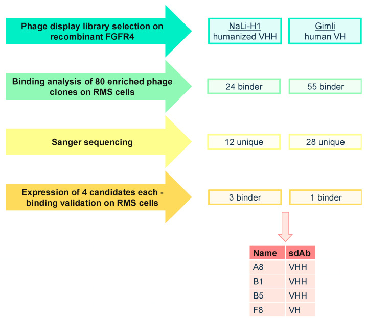
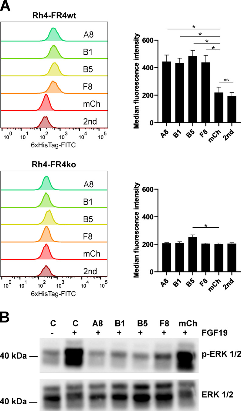
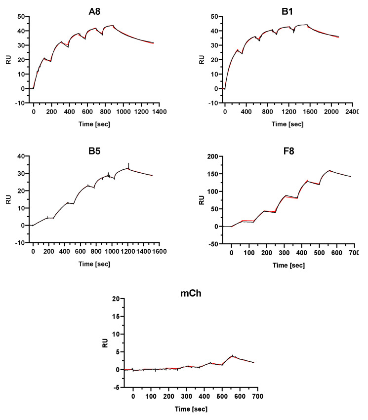
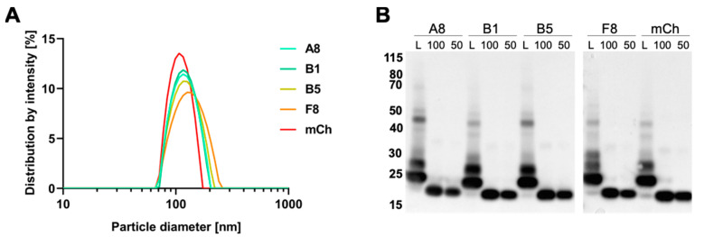
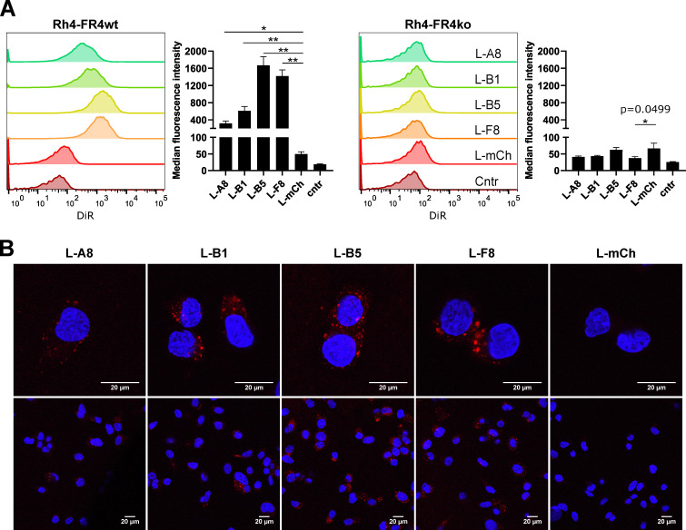
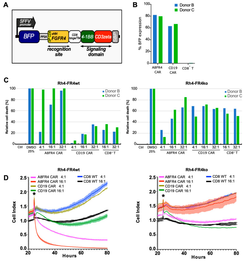
The fibroblast growth factor receptor 4 (FGFR4) is overexpressed in rhabdomyosarcoma (RMS) and represents a promising target for treatments based on specific and efficient antibodies. Despite progress, there is an urgent need for targeted treatment options to improve survival rates, and to limit long-term side effects. From phage display libraries we selected FGFR4-specific single-domain antibodies (sdAb) binding to recombinant FGFR4 and validated them by flow cytometry, surface plasmon resonance, and fluorescence microscopy. The specificity of the selected sdAb was verified on FGFR4-wild type and FGFR4-knock out cells. FGFR4-sdAb were used to decorate vincristine-loaded liposomes and to generate chimeric antigen receptor (CAR) T cells. First, incubation of RMS cells with FGFR4-sdAb revealed that FGFR4-sdAb can block FGF19-FGFR4 signaling via the MAPK pathway and could therefore serve as therapeutics for FGFR4-dependent cancers. Second, FGFR4-targeted vincristine-loaded liposomes bound specifically to RMS cells and were internalized by the receptor, demonstrating the potential for active drug delivery to the tumor. Third, FGFR4-CAR T cells, generated with one sdAb candidate, demonstrated strong and specific cytotoxicity against FGFR4 expressing RMS cells. We selected novel FGFR4-sdAb with high specificity and nano- to picomolar affinities for FGFR4 which have the potential to enable multiple FGFR4-targeted cancer therapy approaches.

Keywords: CAR T cells; FGFR4; rhabdomyosarcoma; single-domain antibody; targeted liposomes.


Conflict of interest statement

N.A., S.M., Z.G., F.P. and M.B. have filed a patent to protect the commercial use of these data. The funders had no role in the design of the study; in the collection, analyses, or interpretation of data; in the writing of the manuscript, or in the decision to publish the results.

Figures

Figure 1
Figure 1
Schematic overview of phage display biopanning and preselection of FGFR4 binding single-domain antibodies (sdAb) sequences. Phage display selection was performed on biotinylated and Dynabeads-bound FGFR4 with two different synthetic sdAb phage display libraries. Enriched phage clones were tested for their binding to cell-surface FGFR4 on Rh4-FR4wt cells resulting in 40 unique binders. Eight sdAb were expressed in E. coli and purified. Of these, recombinant A8, B1, B5 and F8 bound to Rh4-FR4wt but not to Rh4-FR4ko cells.
Figure 2
Figure 2
In vitro binding validation of sdAb. (A) SdAb were tested for their binding selectivity to cell surface FGFR4 by flow cytometry. Histograms show the single-cell living population of each sdAb binding to Rh4-FR4wt versus Rh4-FR4ko cells. Secondary FITC-labeled anti-6xHis-tag antibody alone (2nd) was used as background control and mCherry (mCh) was used as negative control. Median fluorescence intensities (MFI) were determined with FlowJoTM10 software. Statistics: paired t-test, n = 3 independent experiments, mean + SD, * p ≤ 0.05, ns = not significant. (B) Activation assay of FGFR4 in Rh30 cells was performed with recombinant FGF19 and in combination with sdAb. The cells were incubated for 1 h with sdAb at 10 μM (A8, B1, B5, F8, mCh) followed by stimulation of FGFR4 with 50 nM FGF19 for 10 min. Control cells were either not stimulated or stimulated with FGF19 in absence of the sdAb. The cell lysates were analyzed by Western blot with anti-phospho-ERK1/2 antibody. Total Erk1/2 levels are shown as loading control. The complete pictures of the Western blots can be found in Figure S3.
Figure 3
Figure 3
Affinity determination of sdAb to recombinant protein via surface plasmon resonance spectroscopy. Single-cycle kinetics analysis was performed on immobilized FGFR4 through covalent amine binding on the dextran-based sensor chip. The analytes A8, B1, B5, F8 and mCh were injected in 5 different concentrations followed by a dissociation phase. A final dissociation step was added after the last injection step to determine Koff rates for the KD calculations representing binding affinities. The black curves represent the measured data and red curves show the fit analysis (heterogeneous ligand model) performed with the BIAevaluation software.
Figure 4
Figure 4
Characterization of vincristine-loaded targeted liposomes. (A) Size distribution of sdAb-coated liposomes measured by dynamic light scattering. (B) Western blot analysis of coupled sdAb. Liposome suspensions (L) equivalent to 100 ng of sdAb were loaded under reducing and denaturing conditions for gel electrophoresis. Amounts of 100 ng and 50 ng of uncoupled protein were loaded as control. SdAb were detected with an anti-6xHis-tag antibody.
Figure 5
Figure 5
In vitro binding validation of FGFR4 targeting liposomes. (A). Liposomes decorated with FGFR4 targeting sdAb A8, B1, B5 and F8 or mCh (negative control) were tested for their binding selectivity to cell surface FGFR4 by flow cytometry. Adherent cells were incubated for 2 h with 0.5 mM total lipid concentration at 37 °C and 5% CO2. Histograms show the single-cell living population of liposomes binding to Rh4-FR4wt versus Rh4-FR4ko cells. Non-treated cells represent the control populations. Median fluorescence intensities (MFI) are shown. Statistics: paired t-test, n = 3 independent experiments, mean +SD, * p ≤ 0.05, ** p ≤ 0.01. (B) Confocal microscopy analysis of Rh4-FR4wt cells incubated for 2 h at 37 °C and 5% CO2 with sdAb coated fluorescent liposomes. The total lipid concentration was 3 mM. Cells were washed, fixed and mounted with DAPI containing medium. Internalization was visible only in Rh4-FR4wt cells incubated with FGFR4-targeted liposomes.
Figure 6
Figure 6
Cytotoxicity of FGFR4 chimeric antigen receptor (CAR) T cells towards Rhabdomyosarcoma (RMS) cells. (A) Schematic representation of the A8FR4 CAR construct. The CAR is composed of the sdAb A8 with CD8α signal peptide sequence and C-terminal myc-tag followed by the hinge and transmembrane (TM) domains of CD8α. Intracellular signaling domains are 4-1BB and CD3ζ and are followed by a streptavidin binding peptide (SBP). (B) CD8+ T cell transduction efficiencies of donor B and C were determined by flow cytometry analysis of BFP signal. (C) Cytotoxicity was determined by luciferase activity of Rh4 cells co-cultured for 72 h with effector T cells of donors B and C. Relative cell death was highest for Rh4-FR4wt cells incubated with A8FR4 CAR T cells at the indicated effector:target (E:T) cell ratios in both donors. In Rh4-FR4ko cells, non-specific cell killing was observed for the co-cultivation of all CAR T cells and the non-transduced CD8+ T cells. (D) Real-time cell death analysis of Rh4 cells co-cultured with effector T cells from donor B using xCELLigence RTCA DP. A8FR4-CAR T cells showed higher killing activity at the indicated E:T cell ratios in Rh4-FR4wt compared to non-specific CD19 CAR T cells or non-transduced CD8+ T cells. In Rh4-FR4ko cells, no specific cytotoxicity was observed. Horizontal lines within the curves indicate the SD of the duplicate wells used during the assay. The asterisks indicate the time of addition of the effector T cells.

References

    1. Skapek S.X., Ferrari A., Gupta A.A., Lupo P.J., Butler E., Shipley J., Barr F.G., Hawkins D.S. Rhabdomyosarcoma. Nat. Rev. Dis. Prim. 2019;5:1–19. doi: 10.1038/s41572-018-0051-2. - DOI - PMC - PubMed
    1. Hern J.F., Chen L., Chmielecki J., Wei J.S., Patidar R., Rosenberg M., Ambrogio L., Auclair D., Wang J., Song Y.K., et al. Comprehensive Genomic Analysis of Rhabdomyosarcoma Reveals a Landscape of Alterations Affecting a Common Genetic Axis in Fusion-Positive and Fusion-Negative Tumors. Cancer Discov. 2014;4:216–231. doi: 10.1158/2159-8290.cd-13-0639. - DOI - PMC - PubMed
    1. Van Gaal J.C., Van Der Graaf W.T.A., Rikhof B., Van Hoesel Q.G.C.M., Teerenstra S., Suurmeijer A.J.H., Flucke U.E., Loeffen J.L.C.M., Sleijfer S., De Bont E.S.J.M. The impact of age on outcome of embryonal and alveolar rhabdomyosarcoma patients. A multicenter study. Anticancer Res. 2012;32:4485–4497. - PubMed
    1. Sultan I., Qaddoumi I., Yaser S., Rodriguez-Galindo C., Ferrari A. Comparing Adult and Pediatric Rhabdomyosarcoma in the Surveillance, Epidemiology and End Results Program, 1973 to 2005: An Analysis of 2600 Patients. J. Clin. Oncol. 2009;27:3391–3397. doi: 10.1200/JCO.2008.19.7483. - DOI - PubMed
    1. Punyko J.A., Mertens A.C., Gurney J.G., Yasui Y., Donaldson S.S., Rodeberg D.A., Raney R.B., Stovall M., Sklar C.A., Robison L.L., et al. Long-term medical effects of childhood and adolescent rhabdomyosarcoma: A report from the childhood cancer survivor study. Pediatr. Blood Cancer. 2005;44:643–653. doi: 10.1002/pbc.20310. - DOI - PubMed