

Diagnostic Value of Initial Chest CT Findings for the Need of ICU Treatment/Intubation in Patients with COVID-19
- PMID: 33182695
- PMCID: PMC7696816
- DOI: 10.3390/diagnostics10110929
Diagnostic Value of Initial Chest CT Findings for the Need of ICU Treatment/Intubation in Patients with COVID-19
Abstract
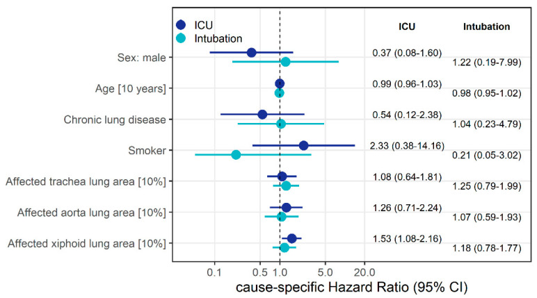
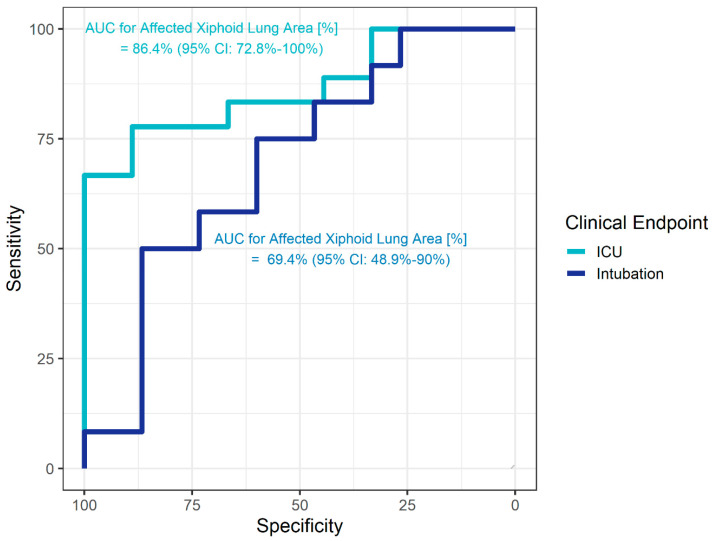
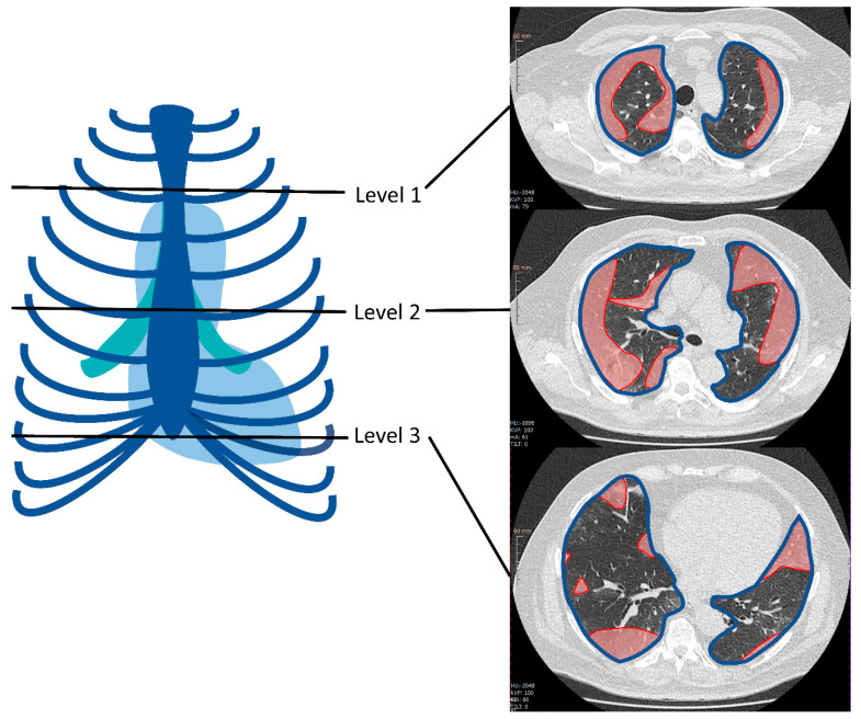
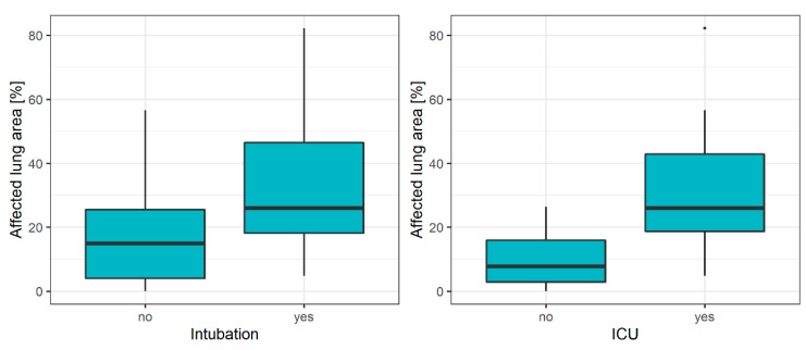
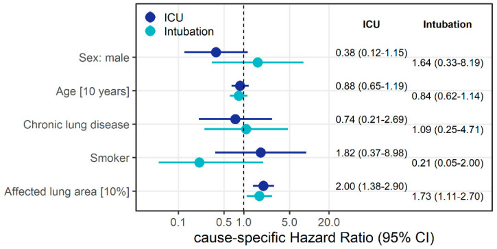
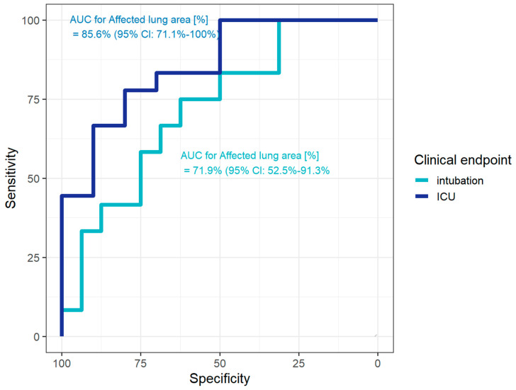
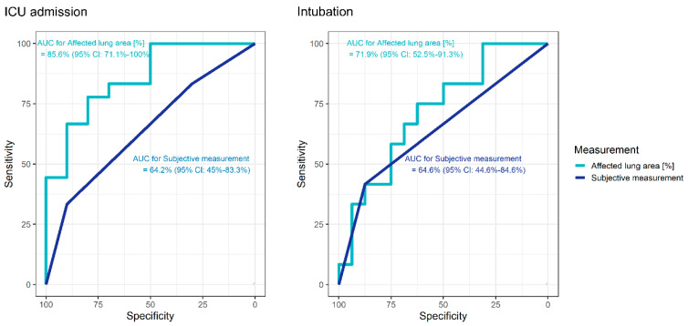
Computed tomography (CT) plays an important role in the diagnosis of COVID-19. The aim of this study was to evaluate a simple, semi-quantitative method that can be used for identifying patients in need of subsequent intensive care unit (ICU) treatment and intubation. We retrospectively analyzed the initial CT scans of 28 patients who tested positive for SARS-CoV-2 at our Level-I center. The extent of lung involvement on CT was classified both subjectively and with a simple semi-quantitative method measuring the affected area at three lung levels. Competing risks Cox regression was used to identify factors associated with the time to ICU admission and intubation. Their potential diagnostic ability was assessed with receiver operating characteristic (ROC)/area under the ROC curves (AUC) analysis. A 10% increase in the affected lung parenchyma area increased the instantaneous risk of intubation (hazard ratio (HR) = 2.00) and the instantaneous risk of ICU admission (HR 1.73). The semi-quantitative measurement outperformed the subjective assessment diagnostic ability (AUC = 85.6% for ICU treatment, 71.9% for intubation). This simple measurement of the involved lung area in initial CT scans of COVID-19 patients may allow early identification of patients in need of ICU treatment/intubation and thus help make optimal use of limited ICU/ventilation resources in hospitals.
Keywords: COVID-19; CT; ICU; SARS-CoV-2; intubation; ventilation.
Conflict of interest statement
The authors declare no conflict of interest.
Figures
Similar articles
-
CT-Based Risk Stratification for Intensive Care Need and Survival in COVID-19 Patients-A Simple Solution.Diagnostics (Basel). 2021 Sep 4;11(9):1616. doi: 10.3390/diagnostics11091616. Diagnostics (Basel). 2021. PMID: 34573957 Free PMC article.
-
3D CT-Inclusive Deep-Learning Model to Predict Mortality, ICU Admittance, and Intubation in COVID-19 Patients.J Digit Imaging. 2023 Apr;36(2):603-616. doi: 10.1007/s10278-022-00734-4. Epub 2022 Nov 30. J Digit Imaging. 2023. PMID: 36450922 Free PMC article.
-
Risk factors associated with mortality in ıntensive care COVID-19 patients: the importance of chest CT score and intubation timing as risk factors.Turk J Med Sci. 2021 Aug 30;51(4):1665-1674. doi: 10.3906/sag-2101-89. Turk J Med Sci. 2021. PMID: 33957728 Free PMC article.
-
Predictors of the chest CT score in COVID-19 patients: a cross-sectional study.Virol J. 2021 Nov 18;18(1):225. doi: 10.1186/s12985-021-01699-6. Virol J. 2021. PMID: 34794467 Free PMC article. Review.
-
Prognostic findings for ICU admission in patients with COVID-19 pneumonia: baseline and follow-up chest CT and the added value of artificial intelligence.Emerg Radiol. 2022 Apr;29(2):243-262. doi: 10.1007/s10140-021-02008-y. Epub 2022 Jan 20. Emerg Radiol. 2022. PMID: 35048222 Free PMC article. Review.
Cited by
-
Treatment of Acute Respiratory Distress Syndrome Caused by COVID-19 with Human Umbilical Cord Mesenchymal Stem Cells.Int J Mol Sci. 2023 Feb 23;24(5):4435. doi: 10.3390/ijms24054435. Int J Mol Sci. 2023. PMID: 36901868 Free PMC article.
-
On the Adoption of Radiomics and Formal Methods for COVID-19 Coronavirus Diagnosis.Diagnostics (Basel). 2021 Feb 12;11(2):293. doi: 10.3390/diagnostics11020293. Diagnostics (Basel). 2021. PMID: 33673394 Free PMC article.
-
Machine learning-based prognostic modeling using clinical data and quantitative radiomic features from chest CT images in COVID-19 patients.Comput Biol Med. 2021 May;132:104304. doi: 10.1016/j.compbiomed.2021.104304. Epub 2021 Mar 3. Comput Biol Med. 2021. PMID: 33691201 Free PMC article.
-
CT-Based Risk Stratification for Intensive Care Need and Survival in COVID-19 Patients-A Simple Solution.Diagnostics (Basel). 2021 Sep 4;11(9):1616. doi: 10.3390/diagnostics11091616. Diagnostics (Basel). 2021. PMID: 34573957 Free PMC article.
-
Dynamic changes in radiological parameters, immune cells, selected miRNAs, and cytokine levels in peripheral blood of patients with severe COVID‑19.Biomed Rep. 2023 Mar 21;18(5):33. doi: 10.3892/br.2023.1615. eCollection 2023 May. Biomed Rep. 2023. PMID: 37034572 Free PMC article.
References
-
- Wu Y.C., Chen C.S., Chan Y.J. Overview of the 2019 Novel Coronavirus (2019-nCoV): The Pathogen of Severe Specific Contagious Pneumonia (SSCP) J. Chin. Med. Assoc. 2020 doi: 10.1097/jcma.0000000000000270. - DOI
-
- WHO . Report of the WHO-China Joint Mission on Coronavirus Disease 2019 (COVID-19) World Health Organization (WHO); Geneva, Switzerland: 2020.
-
- RKI—Robert Koch Institut Sars-Cov-2 Steckbrief Zur Coronavirus-Krankheit-2019 (Covid-19) [(accessed on 13 April 2020)]; Available online: https://www.rki.de/DE/Content/InfAZ/N/Neuartiges_Coronavirus/Steckbrief.....
LinkOut - more resources
Full Text Sources
Miscellaneous
