Review
. 2020 Nov 10;9(11):2455.
doi: 10.3390/cells9112455.

Molecular Evolution, Neurodevelopmental Roles and Clinical Significance of HECT-Type UBE3 E3 Ubiquitin Ligases

Affiliations
Review

Molecular Evolution, Neurodevelopmental Roles and Clinical Significance of HECT-Type UBE3 E3 Ubiquitin Ligases

Mateusz C Ambrozkiewicz et al. Cells. .

Abstract

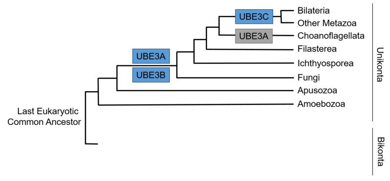
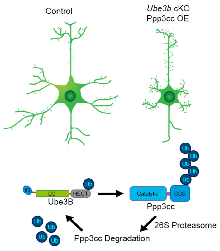
Protein ubiquitination belongs to the best characterized pathways of protein degradation in the cell; however, our current knowledge on its physiological consequences is just the tip of an iceberg. The divergence of enzymatic executors of ubiquitination led to some 600-700 E3 ubiquitin ligases embedded in the human genome. Notably, mutations in around 13% of these genes are causative of severe neurological diseases. Despite this, molecular and cellular context of ubiquitination remains poorly characterized, especially in the developing brain. In this review article, we summarize recent findings on brain-expressed HECT-type E3 UBE3 ligases and their murine orthologues, comprising Angelman syndrome UBE3A, Kaufman oculocerebrofacial syndrome UBE3B and autism spectrum disorder-associated UBE3C. We summarize evolutionary emergence of three UBE3 genes, the biochemistry of UBE3 enzymes, their biology and clinical relevance in brain disorders. Particularly, we highlight that uninterrupted action of UBE3 ligases is a sine qua non for cortical circuit assembly and higher cognitive functions of the neocortex.

Keywords: Angelman syndrome; E3 ubiquitin ligase; Kaufman oculocerebrofacial syndrome; UBE3A; UBE3B; UBE3C; autism spectrum disorder; ubiquitin.


Conflict of interest statement

The authors declare no conflict of interest.

Figures

Figure 1
Figure 1
Sequence alignment of the C-termini of Ube3 ligases (Uniprot accession numbers on the left). Numbers on the left and right indicate the extreme amino acid positions. Highlighted in red is the active site catalytic cysteine. Symbols “*”, “:”, “.”, denote the degree of single residue homology.
Figure 2
Figure 2
Expression levels of Ube3 ligase genes in developing cortex. To plot expression levels of Ube genes, we downloaded the digital gene expression matrix (DGE) provided by Telley et al. [3], in which cells are annotated by the time of collection, and time since FlashTag labelling. We grouped all cells with the same annotation to counts per-gene, and by summing all such values we calculated a “library size” combination of variables, normalizing the counts to control for the number of cells of each type collected. For each gene, we then normalized the count values to the value at the earliest time of collection and “flashtagging”, in order to plot relative change in expression over time.
Figure 3
Figure 3
Simplified pattern of acquisition of Ubiquitin Ligases E3s (UBE3s) across the tree of life of Eucaryota based on [44]. Gains are colored in blue and losses in grey.
Figure 4
Figure 4
Ube3b-mediated Ppp3cc regulation. In the forebrain-specific Ube3b cKO mouse, cortical and hippocampal neurons exhibit simpler, less arborized dendritic trees. This phenotype is mimicked, once neurons overexpress gamma-subunit of calcineurin, Ppp3cc, ubiquitination substrate of Ube3b. LC, low complexity; CBB, calcineurin B and calmodulin binding. PolyUb chain on position on Ppp3cc is randomly placed on the scheme to depict Ube3b-mediated ubiquitination.

References

    1. Rakic P. Evolution of the neocortex: A perspective from developmental biology. Nat. Rev. Neurosci. 2009;10:724–735. doi: 10.1038/nrn2719. - DOI - PMC - PubMed
    1. Nowakowski T.J., Bhaduri A., Pollen A.A., Alvarado B., Mostajo-Radji M.A., Di Lullo E., Haeussler M., Sandoval-Espinosa C., Liu S.J., Velmeshev D., et al. Spatiotemporal gene expression trajectories reveal developmental hierarchies of the human cortex. Science. 2017;358:1318–1323. doi: 10.1126/science.aap8809. - DOI - PMC - PubMed
    1. Telley L., Agirman G., Prados J., Amberg N., Fièvre S., Oberst P., Bartolini G., Vitali I., Cadilhac C., Hippenmeyer S., et al. Temporal patterning of apical progenitors and their daughter neurons in the developing neocortex. Science. 2019:364. doi: 10.1126/science.aav2522. - DOI - PubMed
    1. Dierssen M., Ramakers G.J.A. Dendritic pathology in mental retardation: From molecular genetics to neurobiology. Genes Brain Behav. 2006;5(Suppl. 2):48–60. doi: 10.1111/j.1601-183X.2006.00224.x. - DOI - PubMed
    1. Bliss T.V., Lomo T. Long-lasting potentiation of synaptic transmission in the dentate area of the anaesthetized rabbit following stimulation of the perforant path. J. Physiol. (Lond.) 1973;232:331–356. doi: 10.1113/jphysiol.1973.sp010273. - DOI - PMC - PubMed

Publication types

MeSH terms

Substances

LinkOut - more resources