

Antimicrobial antidegradative dental adhesive preserves restoration-tooth bond
- PMID: 33183773
- PMCID: PMC7704932
- DOI: 10.1016/j.dental.2020.10.017
Antimicrobial antidegradative dental adhesive preserves restoration-tooth bond
Abstract
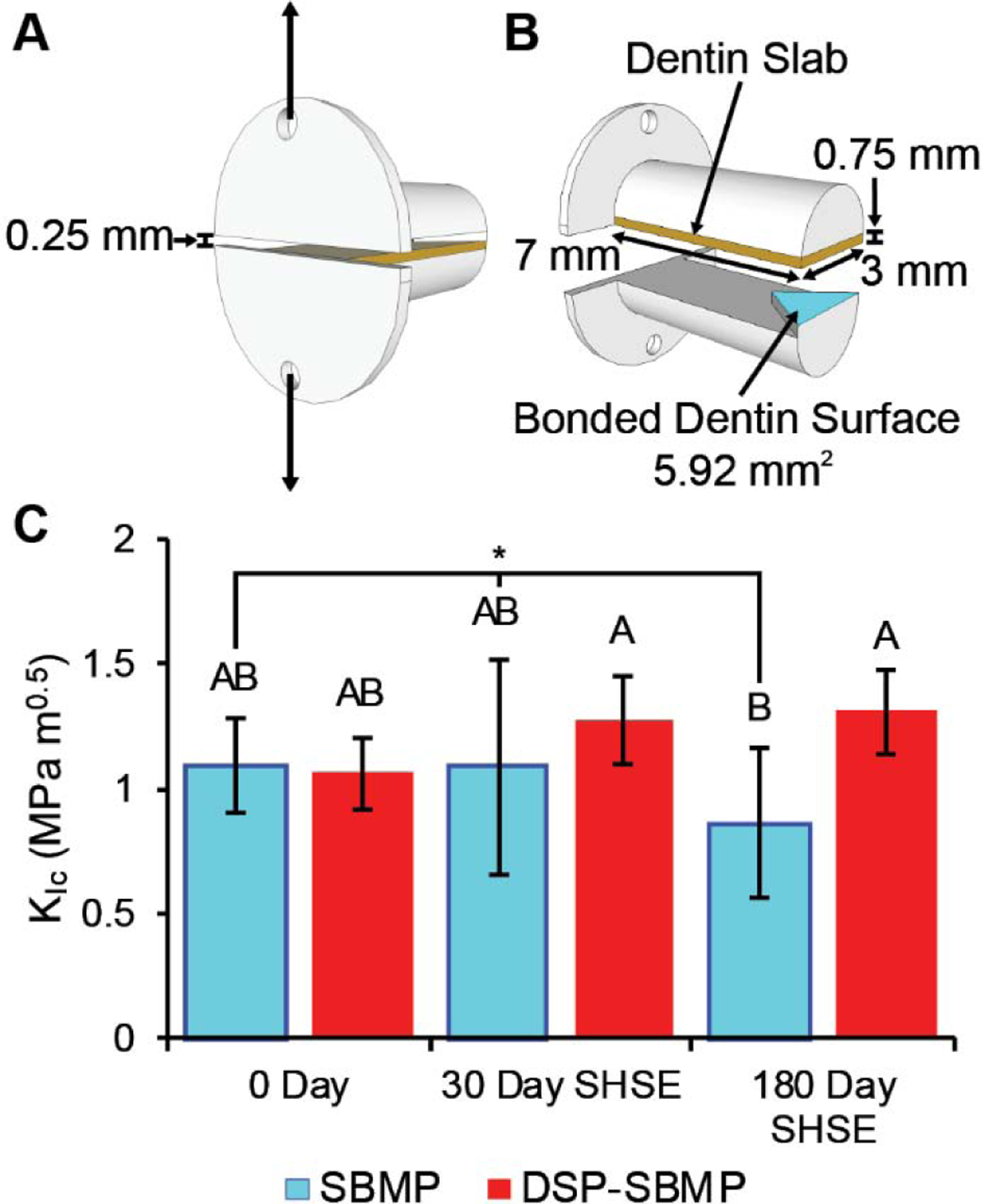
Objective: Assess the ability of an antimicrobial drug-releasing resin adhesive, containing octenidine dihydrochloride (OCT)-silica co-assembled particles (DSPs), to enhance the biostability and preserve the interfacial fracture toughness (FT) of composite restorations bonded to dentin. Enzyme-catalyzed degradation compromises the dental restoration-tooth interface, increasing cariogenic bacterial infiltration. In addition to bacterial ingress inhibition, antimicrobial-releasing adhesives may exhibit direct interfacial biodegradation inhibition as an additional benefit.
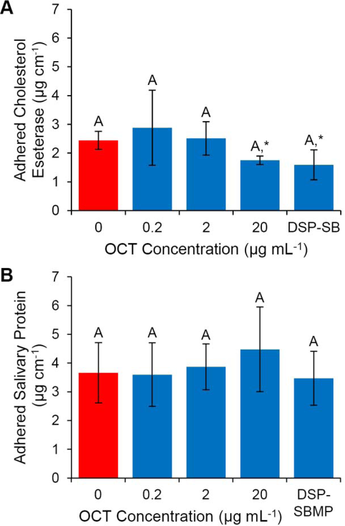
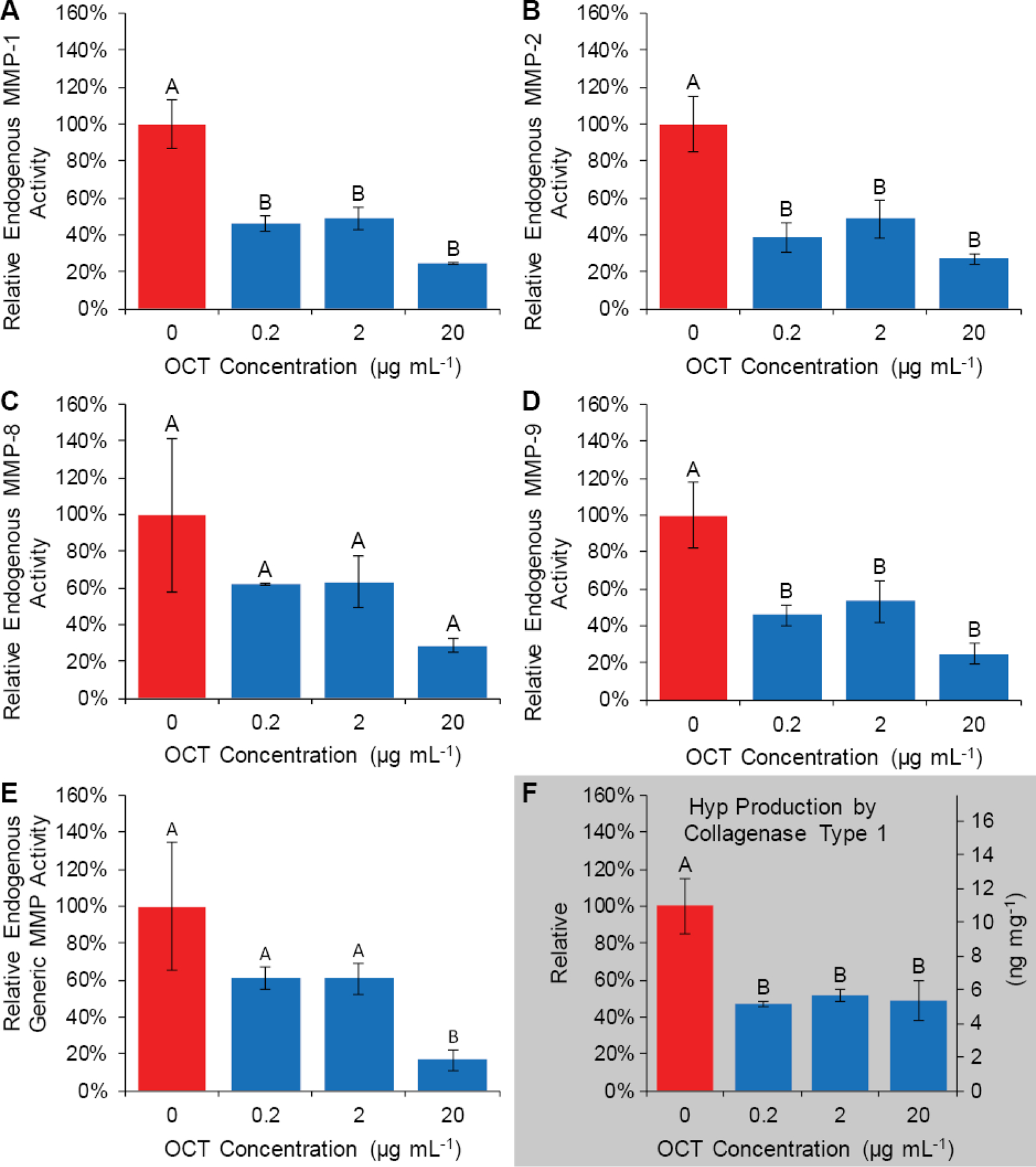
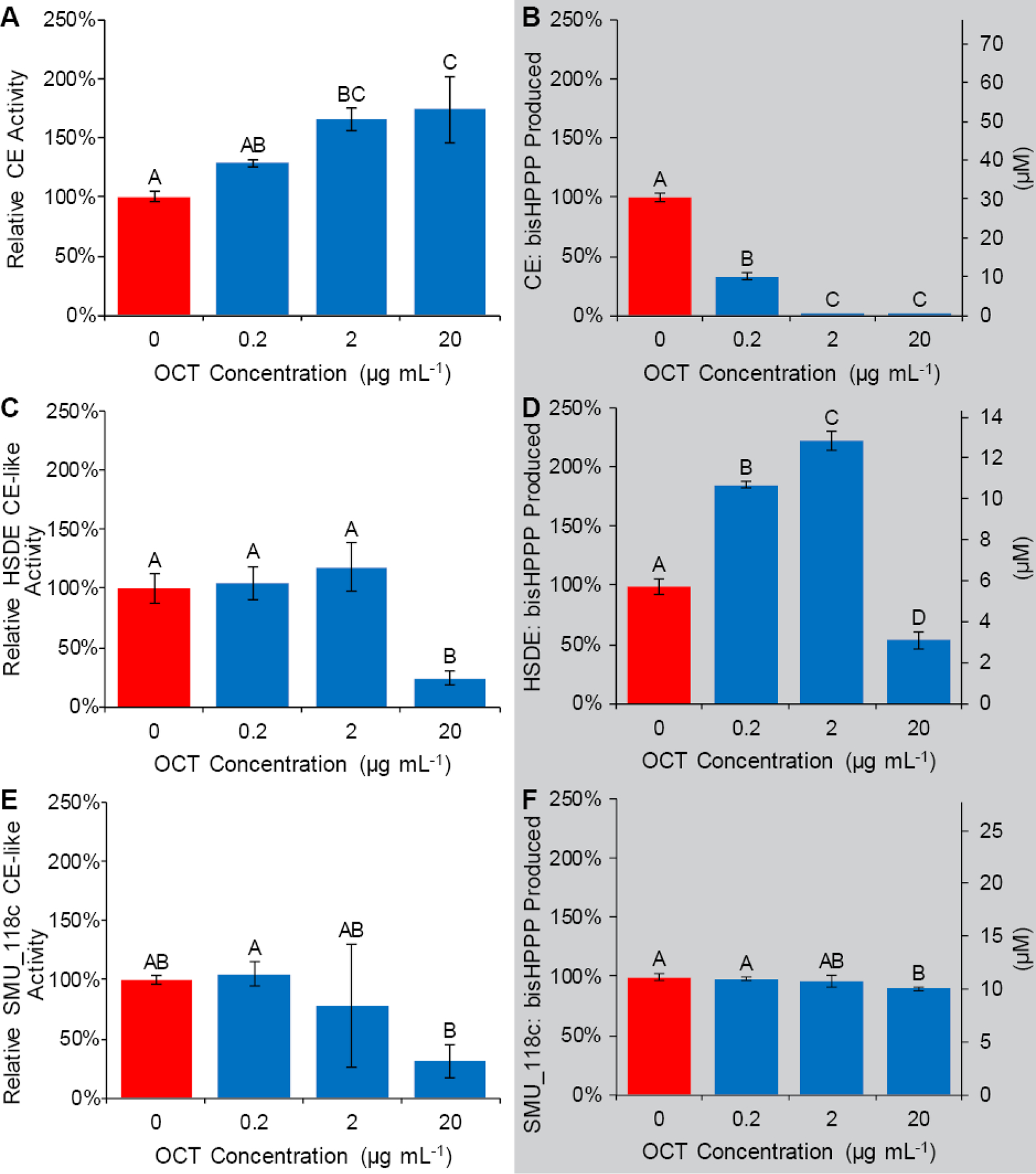
Methods: Mini short-rod restoration bonding specimens with total-etch adhesive with/without 10% wt. DSPs were made. Interfacial fracture toughness (FT) was measured as-manufactured or post-incubation in simulated human salivary esterase (SHSE) for up to 6-months. Effect of OCT on SHSE and whole saliva/bacterial enzyme activity was assessed. Release of OCT outside the restoration interface was assessed.
Results: No deleterious effect of DSPs on initial bonding capacity was observed. Aging specimens in SHSE reduced FT of control but not DSP-adhesive-bonded specimens. OCT inhibited SHSE degradation of adhesive monomer and may inhibit endogenous proteases. OCT inhibited bacterial esterase and collagenase. No endogenous collagen breakdown was detected in the present study. OCT increased human saliva degradative esterase activity below its minimum inhibitory concentration towards S. mutans (MIC), but inhibited degradation above MIC. OCT release outside restoration margins was below detection.
Significance: DSP-adhesive preserves the restoration bond through a secondary enzyme-inhibitory effect of released OCT, which is virtually confined to the restoration interface microgap. Enzyme activity modulation may produce a positive-to-negative feedback switch, by increasing OCT concentration via biodegradation-triggered release to an effective dose, then subsequently slowing degradation and degradation-triggered release.
Keywords: Biodegradation; Dental caries; Enzyme inhibition; Esterase; Interfacial fracture toughness.
Copyright © 2020 The Academy of Dental Materials. Published by Elsevier Inc. All rights reserved.
Conflict of interest statement
Conflict of interest disclosure statement
Authors C. Stewart, B. Hatton and Y. Finer have filed a technology disclosure to the University of Toronto’s Research and Innovation Office, and a PCT application has been submitted (PCT/CA2017/050586 (P1869)) based on the technology and material presented in the manuscript. Author C. Stewart is involved in the commercialization of the technology presented in the manuscript.
Figures
Similar articles
-
Biodegradation of resin-dentin interfaces is dependent on the restorative material, mode of adhesion, esterase or MMP inhibition.Dent Mater. 2018 Sep;34(9):1253-1262. doi: 10.1016/j.dental.2018.05.008. Epub 2018 May 19. Dent Mater. 2018. PMID: 29789163
-
Matrix metalloproteinase inhibitor modulates esterase-catalyzed degradation of resin-dentin interfaces.Dent Mater. 2016 Dec;32(12):1513-1523. doi: 10.1016/j.dental.2016.09.007. Epub 2016 Oct 27. Dent Mater. 2016. PMID: 27692555
-
Human neutrophils compromise the restoration-tooth interface.Acta Biomater. 2020 Nov;117:283-293. doi: 10.1016/j.actbio.2020.09.025. Epub 2020 Sep 17. Acta Biomater. 2020. PMID: 32950724
-
The role of adhesive materials and oral biofilm in the failure of adhesive resin restorations.Am J Dent. 2017 Oct;30(5):285-292. Am J Dent. 2017. PMID: 29178733 Review.
-
Influence of Human and Bacterial Enzymes on Resin Restorations: A Review.J Contemp Dent Pract. 2022 Mar 1;23(3):371-377. J Contemp Dent Pract. 2022. PMID: 35781444 Review.
Cited by
-
Effect of Bioactive and Antimicrobial Nanoparticles on Properties and Applicability of Dental Adhesives.Nanomaterials (Basel). 2022 Nov 1;12(21):3862. doi: 10.3390/nano12213862. Nanomaterials (Basel). 2022. PMID: 36364638 Free PMC article.
-
Anti-Demineralization Effects of Dental Adhesive-Composites on Enamel-Root Dentin Junction.Polymers (Basel). 2021 Sep 29;13(19):3327. doi: 10.3390/polym13193327. Polymers (Basel). 2021. PMID: 34641143 Free PMC article.
-
Streptococcus mutans Proteases Degrade Dentinal Collagen.Dent J (Basel). 2022 Nov 28;10(12):223. doi: 10.3390/dj10120223. Dent J (Basel). 2022. PMID: 36547039 Free PMC article.
-
Numerical Study of the Mechanical Behaviour of Wedge-Shaped Defect Filling Materials.Materials (Basel). 2022 Oct 21;15(20):7387. doi: 10.3390/ma15207387. Materials (Basel). 2022. PMID: 36295452 Free PMC article.
-
Utilizing a degradation prediction pathway system to understand how a novel methacrylate derivative polymer with flipped external ester groups retains physico-mechanical properties following esterase exposure.Dent Mater. 2022 Feb;38(2):251-265. doi: 10.1016/j.dental.2021.12.008. Epub 2021 Dec 18. Dent Mater. 2022. PMID: 34933759 Free PMC article.
References
-
- Kopperud SE, Tveit AB, Gaarden T, Sandvik L, Espelid I. Longevity of posterior dental restorations and reasons for failure. Eur J Oral Sci. 2012;120:539–48. - PubMed
-
- Demarco FF, Correa MB, Cenci MS, Moraes RR, Opdam NJ. Longevity of posterior composite restorations: not only a matter of materials. Dent Mater. 2012;28:87–101. - PubMed
-
- Bernardo M, Luis H, Martin MD, Leroux BG, Rue T, Leitao J, et al. Survival and reasons for failure of amalgam versus composite posterior restorations placed in a randomized clinical trial. Journal of the American Dental Association (1939). 2007;138:775–83. - PubMed
-
- Murray PE, Hafez AA, Smith AJ, Cox CF. Bacterial microleakage and pulp inflammation associated with various restorative materials. Dent Mater. 2002;18:470–8. - PubMed
-
- Soncini JA, Maserejian NN, Trachtenberg F, Tavares M, Hayes C. The longevity of amalgam versus compomer/composite restorations in posterior primary and permanent teeth: findings From the New England Children’s Amalgam Trial. J Am Dent Assoc. 2007;138:763–72. - PubMed
Publication types
MeSH terms
Substances
Grants and funding
LinkOut - more resources
Full Text Sources
Miscellaneous
