Review
. 2021 Jan;31(1):50-61.
doi: 10.1016/j.tcb.2020.10.004. Epub 2020 Nov 9.

Cutting, Amplifying, and Aligning Microtubules with Severing Enzymes

Affiliations
Review

Cutting, Amplifying, and Aligning Microtubules with Severing Enzymes

Yin-Wei Kuo et al. Trends Cell Biol. 2021 Jan.

Abstract

Microtubule-severing enzymes - katanin, spastin, fidgetin - are related AAA-ATPases that cut microtubules into shorter filaments. These proteins, also called severases, are involved in a wide range of cellular processes including cell division, neuronal development, and tissue morphogenesis. Paradoxically, severases can amplify the microtubule cytoskeleton and not just destroy it. Recent work on spastin and katanin has partially resolved this paradox by showing that these enzymes are strong promoters of microtubule growth. Here, we review recent structural and biophysical advances in understanding the molecular mechanisms of severing and growth promotion that provide insight into how severing enzymes shape microtubule networks.

Keywords: microtubule dynamics; microtubule nucleation; severase.


Figures

Figure 1.
Figure 1.. Phylogeny and Structure of Microtubule-Severing Enzymes.
(A) Phylogenetic tree of the meiotic clade AAA-ATPases from model organisms [89]. (B) General architecture of severing enzymes. The predominant microtubule-binding site is located in the microtubule interacting and trafficking (MIT) domain and/or the linker regions. (C) Cryoelectron microscopy structure of a human spastin hexamer bound to a tubulin C-terminal tail (CTT)-mimetic peptide in the presence of the ATP analog ADP-BeFx based on PDB structure 6PEN [33]. The hexamer is not a flat ring. Five protomers of the hexamer are in a right-handed helix, while the sixth protomer (magenta) is in an intermediate position, which closes the ring. (D) Hand-over-hand model of tubulin polypeptide being unfolded by the hexamer (microtubule not to scale). The magenta (apo) subunit moves up to the top of the spiral when binding to ATP (magenta arrow), causing the extraction of an additional two amino acids from the CTT peptide. The yellow subunit is pushed down, leading to ATP hydrolysis, while the cyan subunit subsequently releases ADP and moves up to the position where the apo subunit breaks the spiral.
Figure 2.
Figure 2.. Molecular Mechanism of Microtubule Severing by Severases.
The prevailing model of microtubule severing requires several steps: assembly of hexamers, binding to the tubulin C-terminal tail (CTT), pulling the tubulin polypeptide through the pore, and the generation of lattice defects that eventually lead to microtubule breakage. GTP-tubulin can be added to the defects before a severing event occurs, leading to lattice exchange.
Figure 3.
Figure 3.. Microtubule Length Control by Severing Enzymes.
(A) Inhibition of katanin results in abnormally long spindle length in Xenopus extracts. Adapted from [65]. Top: control spindle; bottom: inhibition of katanin by addition of katanin antibodies. (B) Theoretical microtubule length distributions in the presence of severing [50]. Increased severing activity leads to shorter and more uniform microtubule lengths. Broken lines indicate the average length in each case.
Figure 4.
Figure 4.. Maintenance and Reorientation of Cortical Microtubule Arrays by Selective Severing.
(A) Stimulation by blue light induces cortical microtubule reorientation in Arabidopsis hypocotyl cells. Adapted from [16]. This phototropic response requires the severing activity of katanin. Wild-type (WT) (top) cortical microtubule arrays are highly parallel and reorient to ~90° after the illumination with blue light, while mutations in katanin (bottom) abolish the array reorientation. Bars, 5 μm. (B) Model for plant cortical microtubule array reorientation by phototropic signals. Microtubules crossing the original parallel arrays are severed by katanin. The rapid regrowth of severed fragments amplifies the discordant microtubules and gives rise to reorientation in response to blue-light illumination. (C) Model for the maintenance of parallel microtubule arrays by severing. The microtubule that crosses the original parallel microtubules at a large angle is severed, leading to rapid shrinkage and thus a shorter lifetime. The elimination of the discordant microtubules by severing and collision-induced catastrophe (not shown) together promotes order and maintains microtubule arrays.
Figure I.
Figure I.. Severing-Dependent Microtubule Disassembly.
(A,B) Overexpression of spastin in a rat fibroblast fragments the microtubules. Bar, 10 μm. Adapted from [54]. (C) Model scheme of microtubule loss due to severing. The newly created plus end without a GTP cap rapidly depolymerizes. Under low- or no-rescue conditions, severing eventually leads to a decrease of microtubule length, number, and mass.
Figure I.
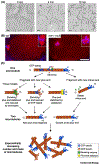
Figure I.. Increase in Microtubule Number and Mass by Severases.
(A) Example of microtubule amplification in vitro. Adapted from [20]. Unlabeled microtubules are visualized by interference reflection microscopy (IRM); the times after addition of ATP and spastin are indicated. Severing of dynamic microtubules by spastin increases the total microtubule number and mass exponentially over time. Bar, 5 μm. (B) Caenorhabditis elegans katanin-null mutant (mei-1) shows a decrease in microtubule (red) number and mass in the embryo. Adapted from [42]. Insets show the magnified meiotic spindles. Bar, 10 μm. (C) Proposed models of severase-dependent microtubule amplification. Severing generates a stable minus end and an unstable plus end. The new plus end can either shrink or be stabilized by plus-end stabilizers. The shrinking plus end can rescue due to its direct stabilization by severases or can rescue when it encounters a GTP island. Abbreviation: WT, wild type.

References

    1. Akhmanova A and Steinmetz MO (2015) Control of microtubule organization and dynamics: two ends in the limelight. Nat. Rev. Mol. Cell Biol 16, 711–726 - PubMed
    1. Goodson HV and Jonasson EM (2018) Microtubules and microtubule-associated proteins. Cold Spring Harb. Perspect. Biol 10, a022608. - PMC - PubMed
    1. Brouhard GJ and Rice LM (2018) Microtubule dynamics: an interplay of biochemistry and mechanics. Nat. Rev. Mol. Cell Biol 19, 451–463 - PMC - PubMed
    1. Howard J and Hyman AA (2007) Microtubule polymerases and depolymerases. Curr. Opin. Cell Biol 19, 31–35 - PubMed
    1. Bowne-Anderson H et al. (2015) Regulation of microtubule growth and catastrophe: unifying theory and experiment. Trends Cell Biol. 25, 769–779 - PMC - PubMed

Publication types