. 2021 Jan-Jun:296:100063.
doi: 10.1074/jbc.RA120.015910. Epub 2020 Nov 22.

An autonomous, but INSIG-modulated, role for the sterol sensing domain in mallostery-regulated ERAD of yeast HMG-CoA reductase

Affiliations

An autonomous, but INSIG-modulated, role for the sterol sensing domain in mallostery-regulated ERAD of yeast HMG-CoA reductase

Margaret A Wangeline et al. J Biol Chem. 2021 Jan-Jun.

Abstract

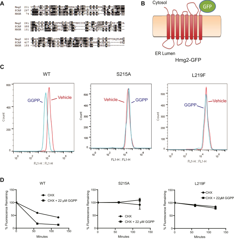
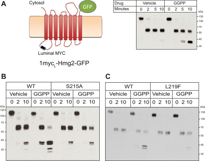
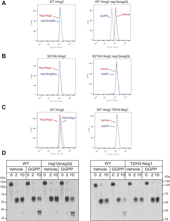
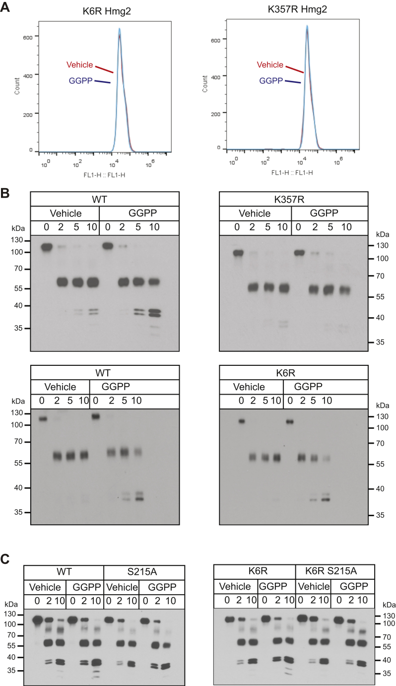
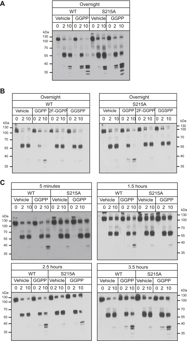
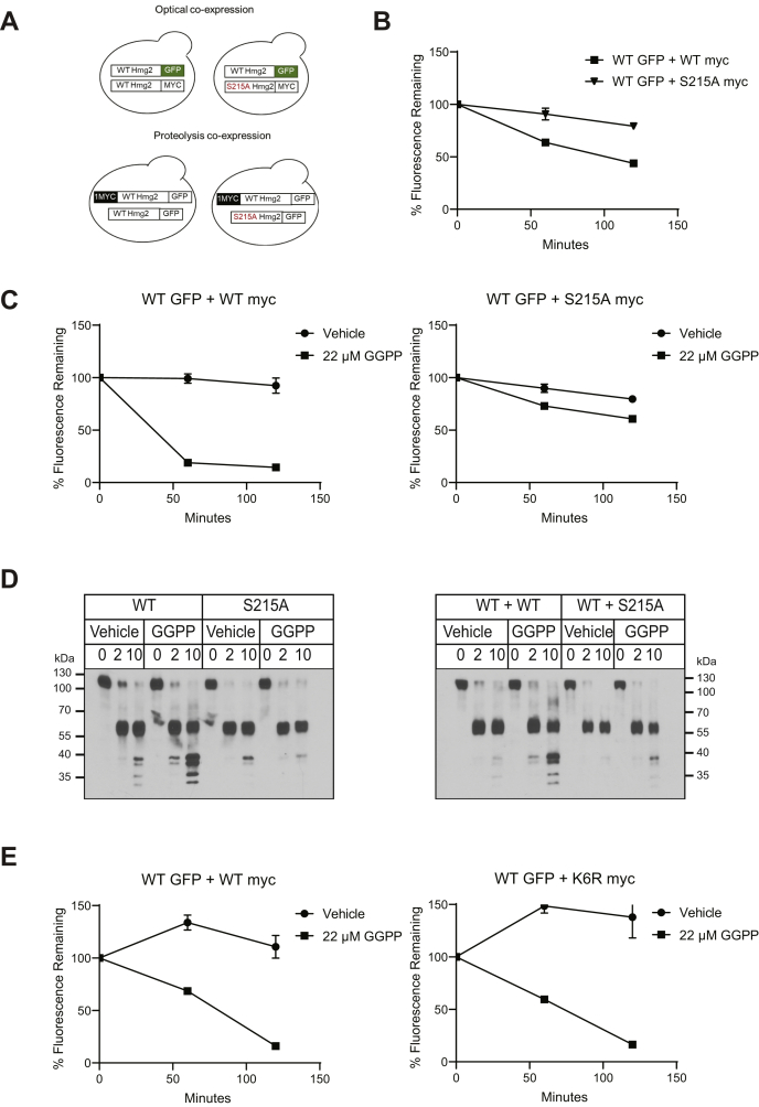
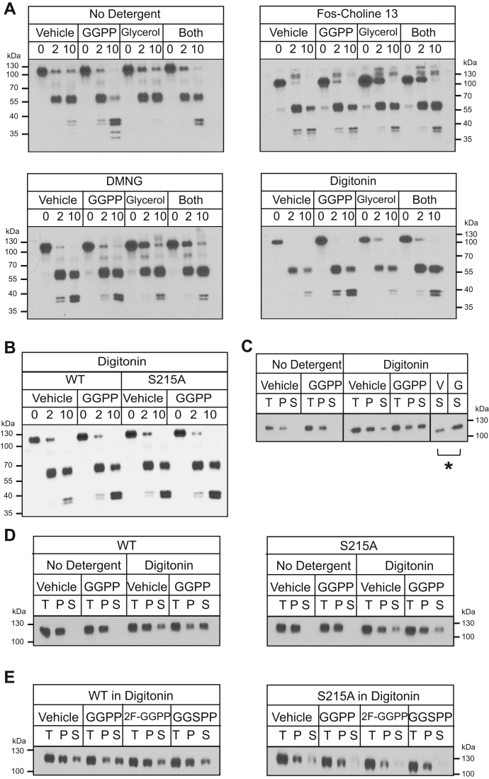
HMG-CoA reductase (HMGR) undergoes feedback-regulated degradation as part of sterol pathway control. Degradation of the yeast HMGR isozyme Hmg2 is controlled by the sterol pathway intermediate GGPP, which causes misfolding of Hmg2, leading to degradation by the HRD pathway; we call this process mallostery. We evaluated the role of the Hmg2 sterol sensing domain (SSD) in mallostery, as well as the involvement of the highly conserved INSIG proteins. We show that the Hmg2 SSD is critical for regulated degradation of Hmg2 and required for mallosteric misfolding of GGPP as studied by in vitro limited proteolysis. The Hmg2 SSD functions independently of conserved yeast INSIG proteins, but its function was modulated by INSIG, thus imposing a second layer of control on Hmg2 regulation. Mutant analyses indicated that SSD-mediated mallostery occurred prior to and independent of HRD-dependent ubiquitination. GGPP-dependent misfolding was still extant but occurred at a much slower rate in the absence of a functional SSD, indicating that the SSD facilitates a physiologically useful rate of GGPP response and implying that the SSD is not a binding site for GGPP. Nonfunctional SSD mutants allowed us to test the importance of Hmg2 quaternary structure in mallostery: a nonresponsive Hmg2 SSD mutant strongly suppressed regulation of a coexpressed, normal Hmg2. Finally, we have found that GGPP-regulated misfolding occurred in detergent-solubilized Hmg2, a feature that will allow next-level analysis of the mechanism of this novel tactic of ligand-regulated misfolding.

Keywords: ER quality control; HMG-CoA reductase; HRD pathway; cholesterol regulation; endoplasmic-reticulum-associated protein degradation (ERAD); mallostery; protein misfolding; sterol sensing domain (SSD); ubiquitin; ubiquitin-proteasome system.


Conflict of interest statement

Conflict of interest The authors declare that they have no conflicts of interest with the contents of this article.

Figures

Figure 1
Figure 1
The Hmg2 sterol sensing domain (SSD) was required for GGPP-regulated degradation. A, amino acid sequence alignment of S. cerevisiae Hmg2 and H. sapiens SCAP and HMGR containing the region of the SSD. Similarities are highlighted in gray, and identities are highlighted in black. Asterisks show the conserved residues S215 and L219. B, schematic of the optical reporter Hmg2-GFP. The transmembrane region of Hmg2 is fused to GFP. (Cartoon originally appeared in (18) and used with permission.). C, SSD mutations rendered Hmg2 more stable in vivo. Histograms of Hmg2-GFP, wild-type, or with the mutations S215A and L219F, as indicated, at steady state (red) or after 2 h of cycloheximide treatment (blue). D, stabilized SSD mutations are GGPP insensitive. Cycloheximide chase of wild-type and the S215A or L219F SSD mutants of Hmg2-GFP, in which CHX is added at time 0, and subsequent levels were measured by flow cytometry, in the presence of vehicle (solid circles) or 22 μM GGPP (solid squares). Error bars are SEM.
Figure 2
Figure 2
The SSD is required for GGPP-mediated mallosteric misfolding in vitro. A, left, schematic of the luminally tagged 1mycL-Hmg2-GFP reporter. A myc tag is inserted into the first luminal loop of Hmg2, and the Hmg2 transmembrane region is fused to GFP. (Cartoon first appeared in (18); used by permission) ER/Golgi microsomes were isolated from strains expressing 1mycL-Hmg2-GFP, treated with vehicle or 22 μM GGPP, and subjected to proteolysis with trypsin. Addition of GGPP accelerates the rate of proteolysis approximately fivefold. B and C, stabilizing SSD mutations blocked GGPP-induced misfolding. Microsomes isolated from strains expressing wild-type or S215A 1mycL-Hmg2-GFP (B), or L219F 1mycL-Hmg2-GFP (C), were treated with vehicle or GGPP prior to proteolysis and then treated with trypsin for the indicated times followed by immunoblotting for the luminal myc tag. GGPP induced misfolding and increased proteolysis in the wild-type protein, but was essentially ineffective.
Figure 3
Figure 3
GGPP-regulated Hmg2 mallosteric misfolding did not require INSIG proteins and was inhibited by excess Nsg1. A absence of yeast INSIGS Nsg1 and Nsg2 did not affect regulated degradation of Hmg2-GFP. Left panel, steady-state levels of Hmg2-GFP in a strain with wild-type INSIG proteins (red curve) or an otherwise identical strain with missing both INSIG genes by virtue of nsg1Δnsg2Δ double null (blue curve). Right panel: in the INSIG double null, 22 μM GGPP treatment caused the expected drop in steady-state Hmg2-GFP levels (blue curve) compared with vehicle (red curve). B, deletion of yeast INSIGs did not alter the stability or GGPP nonresponsiveness of the S215A mutant. Left panel, S215A Hmg2-GFP levels in a wild-type (red curve) or nsg1Δnsg2Δ double null strain (blue curve). Right panel, lack of GGPP responsiveness of S215A mutant in an nsg1Δnsg2Δ strain treated with vehicle (red) or 22 μM GGPP (blue) for 1 h. The block in regulation of the S215A mutant was not affected by INSIG deletion. C, overexpression of Nsg1 increased Hmg2-GFP levels and prevented GGPP-induced degradation. Left, wild-type Hmg2-GFP expressed in a wild-type strain (red) or a strain producing Nsg1 from the strong TDH3 promoter (blue) was evaluated for steady-state fluorescence by flow cytometry. Right, cells expressing Hmg2-GFP in an identical strain expressing Nsg1 from the strong TDH3 promoter were treated with vehicle (red) or 22 μM GGPP (blue) and evaluated for steady-state fluorescence by flow cytometry for 1 h. D, GGPP-induced in vitro misfolding of Hmg2-GFP did not require INSIGs, but was blocked by strong Nsg1 coexpression. Left, in vitro proteolysis of 1mycL-Hmg2-GFP expressed in wild-type and nsg1Δnsg2Δ yeast. 22 μM GGPP induced misfolding and increased the rate of proteolysis in both backgrounds. Right, in vitro proteolysis of 1mycL-Hmg2-GFP expressed in a strain wild-type for INSIGs and a strain expressing Nsg1 from the strong TDH3 promoter. Co-overexpression of Nsg1 blocked GGPP-induced misfolding.
Figure 4
Figure 4
Separate sequence determinants of misfolding and degradation.A, the two lysine-to-arginine mutations, K6R and K357R each rendered Hmg2-GFP insensitive to the degradation signal GGPP. Strains expressing each mutant were treated with 22 μM GGPP (blue curves) for 2 h and then subjected to flow cytometery. Unlike wild-type Hmg2-GFP (Fig. 1C), GGPP did not affect K6R or K357R Hmg2-GFP levels compared with vehicle treatment (red curves). B, the mutation K357R blocked GGPP-induced misfolding in vitro, but the K6R mutation did not. Microsomes expressing wild-type, K357R, and K6R 1mycL-Hmg2-GFP were isolated and treated with vehicle or 22 μM GGPP prior to proteolysis as in earlier figures. GGPP did not cause misfolding and increased proteolysis in the K357R mutant (top panels). Conversely, K6R 1mycL-Hmg2-GFP responded to GGPP like the wild-type protein (bottom panels). C, in-cis epistasis of the K6R mutation and the SSD mutation S215A. Both wild-type and K6R 1mycL-Hmg2-GFP misfolded upon 22 μM GGPP treatment, whereas the K6R S215A double mutation did not respond to GGPP treatment.
Figure 5
Figure 5
GGPP caused mallosteric misfolding of the stable SSD mutant S215A at high concentrations when treated for long time courses. A, overnight GGPP treatment caused in vitro misfolding of S215A Hmg2. Western blots of in vitro proteolysis performed on membranes from cells expressing wild-type or S215A 1mycL-Hmg2-GFP and incubated with vehicle or 22 μM GGPP overnight (15 h). B, GGPP-mediated, long time course misfolding of S215A maintained the high GGPP structural specificity observed in WT Hmg2. Specificity of overnight misfolding of wild-type or S215A 1mycL-Hmg2-GFP was tested by treatment with 22 μM of the close analogs of GGPP, 2-fluouro GGPP (2F-GGPP), and s-thiolo GGPP (GGSPP), shown to be inactive in more canonical rapid structural changes in wild-type Hmg2. Neither caused misfolding of either wild-type or S215A Hmg2 at the high concentrations and long time courses employed in this experiment. C, time course of wild-type and S215A GGPP-induced misfolding. Wild-type 1mycL-Hmg2-GFP misfolded in response to GGPP more quickly than can be measured, within 5 min. S215A 1mycL-Hmg2-GFP began to misfold in response to GGPP after approximately 2.5 h of treatment.
Figure 6
Figure 6
Trans effects of mutated SSDs on regulated misfolding. A, cartoon showing experimental setup for coexpression experiments. For optical experiments, WT Hmg2-GFP was coexpressed alongside a nonfluorescent, myc-tagged mutant S215A or WT (control) copy (top). For proteolysis experiments, WT 1mycL-Hmg2-GFP was coexpressed alongside a GFP-tagged, without myc, mutant S215A Hmg2-GFP or WT (control) (bottom). B, coexpression of dark (nonfluorescent), myc-tagged, nonmallosteric mutant S215A Hmg2 strongly stabilized wild-type Hmg2-GFP. Wild-type Hmg2-GFP was expressed along with dark wild-type Hmg2-myc (filled squares) or S215A Hmg2-myc (filled triangles). In a cycloheximide chase, the dark S215A Hmg2-myc slowed the degradation of wild-type Hmg2-GFP when compared with a strain coexpressing wild-type Hmg2-myc. Error bars are SEM. C, coexpression of the dark S215A Hmg2-myc inhibited the response of wild-type Hmg2-GFP to GGPP. Wild-type Hmg2-GFP was coexpressed with wild-type Hmg2-myc (left) or S215A Hmg2-myc (right). Coexpression of the mutated Hmg2-myc, which cannot misfold, but not the wild-type Hmg2-myc, partially blocked GGPP-induced degradation of the wild-type Hmg2-GFP. Filled circles show vehicle control, and filled squares show GGPP treatment. Error bars are SEM. D, coexpression of a nonmallosteric SSD mutant Hmg2-GFP strongly blocked GGPP-induced misfolding of wild-type 1mycL-Hmg2-GFP in vitro. Left, 22 μM GGPP induced misfolding and caused increased proteolytic cleavage in wild-type but not S215A 1mycL-Hmg2-GFP. Right, coexpression of wild-type Hmg2-GFP without a myc tag did not interfere proteolysis of wild-type 1mycL-Hmg2-GFP. However, coexpression of S215A Hmg2-GFP with no myc tag attenuated GGPP-induced misfolding of the wild-type 1mycL-Hmg2-GFP copy. E, coexpression of the highly stable but still mallosteric K6R Hmg2-myc allowed normal Hmg2-GFP regulation. Cells expressing Hmg2-GFP and coexpressing a dark wild-type Hmg2-myc (left) or K6R Hmg2-myc (right) were treated with vehicle (filled circles) or GGPP (filled squares), and Hmg2-GFP levels were assayed by flow cytometry. The K6R Hmg2-myc did not block regulation of the wild-type Hmg2-GFP. Error bars are SEM.
Figure 7
Figure 7
In vitro proteolysis assay of micellar 1mycL-Hmg2-GFP is intact, and responses to glycerol and GGPP. A, the nonionic detergents fos-choline 13, decyl maltose neopentyl glycol (DMNG), and digitonin allowed the time-dependent proteolytic cleavage pattern of 1mycL-Hmg2-GFP in solution. Microsomes from cells expressing 1mycL-Hmg2-GFP were isolated as previously and left unsolubilized (top left) or subjected to solubilization with fos-choline 13 (top right), DMNG (bottom left), or digitonin (bottom right) as described in text. For the no detergent condition, the microsome pellet was used; for the three detergent conditions, solubilized microsomes were clarified by ultracentrifugation, and the supernatant was subjected to proteolysis. In fos-choline 13, DMNG, and digitonin, the 1mycL-Hmg2-GFP myc tag remained intact during proteolysis. 1mycL-Hmg2-GFP remained responsive to the action of the chemical chaperone glycerol in all three detergents. Furthermore, 1mycL-Hmg2-GFP solubilized in digitonin remained responsive to GGPP in vitro. B, digitonin solubilization preserved the SSD requirement for 1mycL-Hmg2-GFP misfolding. When solubilized with digitonin, proteolysis of wild-type 1mycL-Hmg2-GFP increased in response to GGPP treatment, but proteolysis of S215A 1mycL-Hmg2-GFP did not. C, GGPP treatment during solubilization increased the solubility of 1mycL-Hmg2-GFP. Left, when not solubilized and subjected to centrifugation, 1mycL-Hmg2-GFP was present only in the pellet when detected by western blotting for the myc tag. GGPP did not affect 1mycL-Hmg2-GFP fractionation in unsolubilized microsomes. Right, when microsomes were solubilized with digitonin, 1mycL-Hmg2-GFP was present in both pellet and supernatant fractions. Treatment of microsomes with 22 μM GGPP during solubilization increased the amount of 1mycL-Hmg2-GFP in the supernatant fraction. Far right, comparison of the amount of 1mycL-Hmg2-GFP in the supernatant when treated with vehicle versus GGPP. ∗p ≤ 0.05. D, the solubilization effect of added GGPP was SSD-dependent. Left, treating preparations with GGPP during solubilization increased the amount of wild-type 1mycL-Hmg2-GFP detectable in the supernatant. However, the solubility of S215A 1mycL-Hmg2-GFP was not affected by GGPP. E, the increase in solubilization was specific for GGPP. Treating preparations with the close analogues of GGPP, 2-fluoro-GGPP (2F-GGPP), or s-thiolo GGPP (GGSPP) did not increase the amount of 1mycL-Hmg2-GFP detectable in the supernatant.

Similar articles

Cited by

References

    1. Needham P.G., Brodsky J.L. How early studies on secreted and membrane protein quality control gave rise to the ER associated degradation (ERAD) pathway: the early history of ERAD. Biochim. Biophys. Acta. 2013;1833:2447–2457. - PMC - PubMed
    1. Mehnert M., Sommer T., Jarosch E. ERAD ubiquitin ligases. BioEssays. 2010;32:905–913. - PubMed
    1. Smith M.H., Ploegh H.L., Weissman J.S. Road to ruin: targeting proteins for degradation in the endoplasmic reticulum. Science. 2011;334:1086–1090. - PMC - PubMed
    1. Mehrtash A.B., Hochstrasser M. Ubiquitin-dependent protein degradation at the endoplasmic reticulum and nuclear envelope. Semin. Cell Dev. Biol. 2019;93:111–124. - PMC - PubMed
    1. Hampton R.Y., Rine J. Regulated degradation of HMG-CoA reductase, an integral membrane protein of the endoplasmic reticulum, in yeast. J. Cell Biol. 1994;125:299–312. - PMC - PubMed

Publication types

MeSH terms

Substances