

Crystal structure of a guanine nucleotide exchange factor encoded by the scrub typhus pathogen Orientia tsutsugamushi
- PMID: 33184172
- PMCID: PMC7720168
- DOI: 10.1073/pnas.2018163117
Crystal structure of a guanine nucleotide exchange factor encoded by the scrub typhus pathogen Orientia tsutsugamushi
Abstract
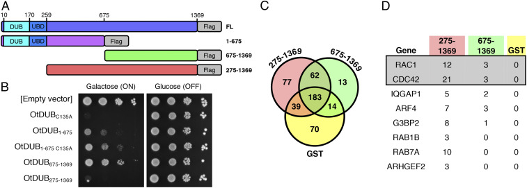
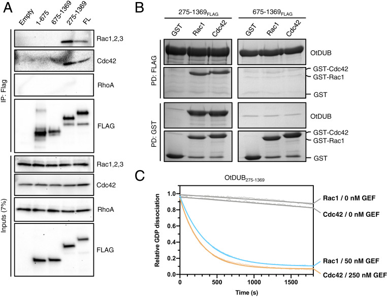
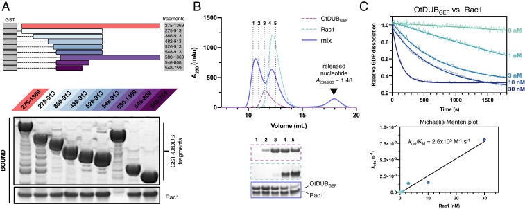
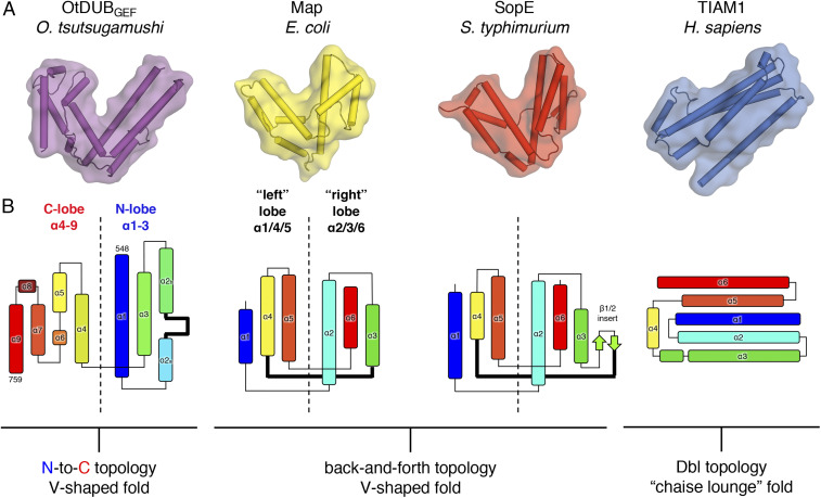
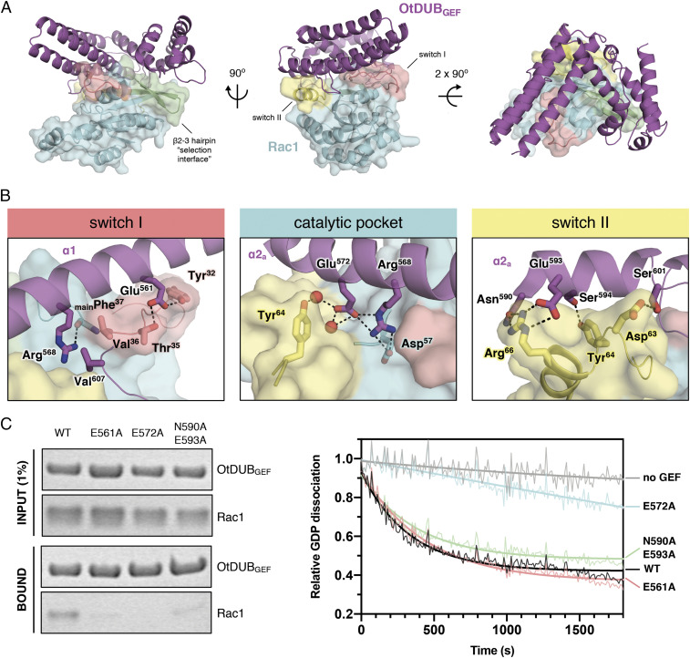
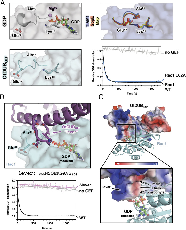
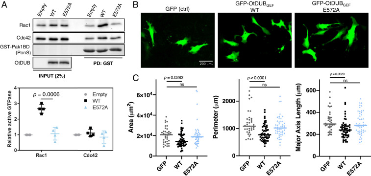
Rho family GTPases regulate an array of cellular processes and are often modulated by pathogens to promote infection. Here, we identify a cryptic guanine nucleotide exchange factor (GEF) domain in the OtDUB protein encoded by the pathogenic bacterium Orientia tsutsugamushi A proteomics-based OtDUB interaction screen identified numerous potential host interactors, including the Rho GTPases Rac1 and Cdc42. We discovered a domain in OtDUB with Rac1/Cdc42 GEF activity (OtDUBGEF), with higher activity toward Rac1 in vitro. While this GEF bears no obvious sequence similarity to known GEFs, crystal structures of OtDUBGEF alone (3.0 Å) and complexed with Rac1 (1.7 Å) reveal striking convergent evolution, with a unique topology, on a V-shaped bacterial GEF fold shared with other bacterial GEF domains. Structure-guided mutational analyses identified residues critical for activity and a mechanism for nucleotide displacement. Ectopic expression of OtDUB activates Rac1 preferentially in cells, and expression of the OtDUBGEF alone alters cell morphology. Cumulatively, this work reveals a bacterial GEF within the multifunctional OtDUB that co-opts host Rac1 signaling to induce changes in cytoskeletal structure.
Keywords: Orientia tsutsugamushi; Rac1; X-ray crystallography; guanine nucleotide exchange factor; scrub typhus.
Conflict of interest statement
The authors declare no competing interest.
Figures
Similar articles
-
Orientia tsutsugamushi OtDUB Is Expressed and Interacts with Adaptor Protein Complexes during Infection.Infect Immun. 2022 Dec 15;90(12):e0046922. doi: 10.1128/iai.00469-22. Epub 2022 Nov 14. Infect Immun. 2022. PMID: 36374099 Free PMC article.
-
A deubiquitylase with an unusually high-affinity ubiquitin-binding domain from the scrub typhus pathogen Orientia tsutsugamushi.Nat Commun. 2020 May 11;11(1):2343. doi: 10.1038/s41467-020-15985-4. Nat Commun. 2020. PMID: 32393759 Free PMC article.
-
Trp(56) of rac1 specifies interaction with a subset of guanine nucleotide exchange factors.J Biol Chem. 2001 Dec 14;276(50):47530-41. doi: 10.1074/jbc.M108865200. Epub 2001 Oct 10. J Biol Chem. 2001. PMID: 11595749
-
The guanine nucleotide exchange factor Tiam1: a Janus-faced molecule in cellular signaling.Cell Signal. 2014 Mar;26(3):483-91. doi: 10.1016/j.cellsig.2013.11.034. Epub 2013 Dec 2. Cell Signal. 2014. PMID: 24308970 Review.
-
Structural biology of DOCK-family guanine nucleotide exchange factors.FEBS Lett. 2023 Mar;597(6):794-810. doi: 10.1002/1873-3468.14523. Epub 2022 Nov 4. FEBS Lett. 2023. PMID: 36271211 Free PMC article. Review.
Cited by
-
Autoinhibition of the GEF activity of cytoskeletal regulatory protein Trio is disrupted in neurodevelopmental disorder-related genetic variants.J Biol Chem. 2022 Sep;298(9):102361. doi: 10.1016/j.jbc.2022.102361. Epub 2022 Aug 10. J Biol Chem. 2022. PMID: 35963430 Free PMC article.
-
Climate drives the spatiotemporal dynamics of scrub typhus in China.Glob Chang Biol. 2022 Nov;28(22):6618-6628. doi: 10.1111/gcb.16395. Epub 2022 Sep 2. Glob Chang Biol. 2022. PMID: 36056457 Free PMC article.
-
Metagenome diversity illuminates origins of pathogen effectors.bioRxiv [Preprint]. 2023 Feb 27:2023.02.26.530123. doi: 10.1101/2023.02.26.530123. bioRxiv. 2023. Update in: mBio. 2024 May 8;15(5):e0075923. doi: 10.1128/mbio.00759-23. PMID: 36909625 Free PMC article. Updated. Preprint.
-
OtDUB from the Human Pathogen Orientia tsutsugamushi Modulates Host Membrane Trafficking by Multiple Mechanisms.Mol Cell Biol. 2022 Jul 21;42(7):e0007122. doi: 10.1128/mcb.00071-22. Epub 2022 Jun 21. Mol Cell Biol. 2022. PMID: 35727026 Free PMC article.
-
Metagenome diversity illuminates the origins of pathogen effectors.mBio. 2024 May 8;15(5):e0075923. doi: 10.1128/mbio.00759-23. Epub 2024 Apr 2. mBio. 2024. PMID: 38564675 Free PMC article.
References
-
- Burridge K., Wennerberg K., Rho and Rac take center stage. Cell 116, 167–179 (2004). - PubMed
-
- Jaffe A. B., Hall A., Rho GTPases: Biochemistry and biology. Annu. Rev. Cell Dev. Biol. 21, 247–269 (2005). - PubMed
-
- Vetter I. R., Wittinghofer A., The guanine nucleotide-binding switch in three dimensions. 294, 1299–1304 (2001). - PubMed
-
- Zhang B., Zhang Y., Wang Z., Zheng Y., The role of Mg2+ cofactor in the guanine nucleotide exchange and GTP hydrolysis reactions of Rho family GTP-binding proteins. J. Biol. Chem. 275, 25299–25307 (2000). - PubMed
Publication types
MeSH terms
Substances
Grants and funding
LinkOut - more resources
Full Text Sources
Research Materials
Miscellaneous
