. 2021 Feb;27(2):133-150.
doi: 10.1261/rna.077123.120. Epub 2020 Nov 12.

Expansion segments in bacterial and archaeal 5S ribosomal RNAs

Affiliations

Expansion segments in bacterial and archaeal 5S ribosomal RNAs

Victor G Stepanov et al. RNA. 2021 Feb.

Abstract

The large ribosomal RNAs of eukaryotes frequently contain expansion sequences that add to the size of the rRNAs but do not affect their overall structural layout and are compatible with major ribosomal function as an mRNA translation machine. The expansion of prokaryotic ribosomal RNAs is much less explored. In order to obtain more insight into the structural variability of these conserved molecules, we herein report the results of a comprehensive search for the expansion sequences in prokaryotic 5S rRNAs. Overall, 89 expanded 5S rRNAs of 15 structural types were identified in 15 archaeal and 36 bacterial genomes. Expansion segments ranging in length from 13 to 109 residues were found to be distributed among 17 insertion sites. The strains harboring the expanded 5S rRNAs belong to the bacterial orders Clostridiales, Halanaerobiales, Thermoanaerobacterales, and Alteromonadales as well as the archael order Halobacterales When several copies of a 5S rRNA gene are present in a genome, the expanded versions may coexist with normal 5S rRNA genes. The insertion sequences are typically capable of forming extended helices, which do not seemingly interfere with folding of the conserved core. The expanded 5S rRNAs have largely been overlooked in 5S rRNA databases.

Keywords: 5S rRNA; archaea; bacteria; expansion segment; ribosome.


Figures

FIGURE 1.
FIGURE 1.
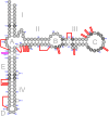
Generalized secondary structure model of prokaryotic 5S rRNA with indicated locations of expansion segments. Universally conserved nucleotides are shown as white characters in black filled circles. Dashed open circles represent nucleotides unique to archaeal 5S rRNAs. Dashed open diamonds represent a unique base pair observed only in 5S rRNAs from the bacterial family Lachnospiraceae. Canonical Watson–Crick bonding is depicted with solid lines. Non-Watson–Crick bonding is presented as small open circles. Dotted lines indicate that the bases may be bound by either Watson–Crick or non-Watson–Crick interactions. Expansion segments are schematically presented as thick red (Bacteria) or magenta (Archaea) lines connecting the flanking residues of the conserved 5S rRNA core.
FIGURE 2.
FIGURE 2.
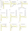
Predicted secondary structures of several representative 5S rRNAs with expansion segments. Nucleotides of the conserved core are highlighted yellow. (A) 5S rRNA from Halococcus morrhuae DSM 1307 (NZ_AOMC01000054.1, 66311–66541). (B) 5S rRNA from Thermoanaerobacterium thermosaccharolyticum DSM 571 (NC_014410.1, 84899–85053). (C) 5S rRNA from Desulfitobacterium dichloroeliminans LMG P-21439 (NC_019903.1, complement [2241573–2241816]). (D) 5S rRNA from Thermacetogenium phaeum DSM 12270 (NC_018870.1, complement [1414325–1414496]). (E) 5S rRNA from Halobacteroides halobius DSM 5150 (NC_019978.1, 20186–20360). (F) 5S rRNA from Halanaerobium sp. DL-01 (NZ_QPJN01000058.1, complement [9–185]). (G) 5S rRNA from Lachnospiraceae bacterium, strain COE1 (NZ_KE159617.1, 786025–786157). (H) 5S rRNA from Pseudoalteromonas piscicida DE2-B (NZ_CP021646.1, complement [2103409–2103538]). (I) 5S rRNA from Desulfallas geothermicus DSM 3669 (NZ_FOYM01000008.1, complement [126967–127097]).
FIGURE 3.
FIGURE 3.
Phylogenetic tree of the archaeal order Halobacteriales. Branches associated with the strains carrying the expanded 5S rRNA genes are highlighted yellow. Branch support values represent branch recovery percentage in 100 jackknife resampling replicates. Scale bar represents the number of nucleotide substitutions per position. The tree was generated using the maximum likelihood algorithm on a 106,995-bp-long concatenated codon-wise alignment of 100 conserved single-copy protein-coding genes shared across all of the 50 strains selected for tree construction. The tree was rooted using Haloferax elongans ATCC BAA-1513 as an outgroup.
FIGURE 4.
FIGURE 4.
Partial phylogenetic tree of the bacterial order Thermoanaerobacterales represented by the related members of families Thermoanaerobacteraceae, Thermoanaerobacterales Family III and Family IV incertae sedis. Branches associated with the strains carrying the expanded 5S rRNA genes are highlighted yellow. The inset shows a scaled-up fragment of the tree. Branch support values represent branch recovery percentage in 100 jackknife resampling replicates. Scale bar represents the number of nucleotide substitutions per position. The tree was generated using the maximum likelihood algorithm on an 80,655-bp-long concatenated codon-wise alignment of 95 conserved single-copy protein-coding genes shared across all of the 37 strains selected for tree construction. The tree was rooted using Anoxybacter fermentans DY22613 as an outgroup.
FIGURE 5.
FIGURE 5.
Phylogenetic tree of the bacterial family Lachnospiraceae. Branches associated with the strains carrying the expanded 5S rRNA genes are highlighted yellow. Branch support values represent branch recovery percentage in 100 jackknife resampling replicates. Scale bar represents the number of nucleotide substitutions per position. The tree was generated using the maximum likelihood algorithm on a 98,322-bp-long concatenated codon-wise alignment of 100 conserved single-copy protein-coding genes shared across all of the 37 strains selected for tree construction. The tree was rooted using Thermacetogenium phaeum DSM 12270 as an outgroup.
FIGURE 6.
FIGURE 6.
Phylogenetic tree of the bacterial order Halanaerobiales. Branches associated with the strains carrying the expanded 5S rRNA genes are highlighted yellow. Branch support values represent branch recovery percentage in 100 jackknife resampling replicates. Scale bar represents the number of nucleotide substitutions per position. The tree was generated using the maximum likelihood algorithm on a 29,271-bp-long concatenated codon-wise alignment of 27 conserved single-copy protein-coding genes shared across all of the 30 strains selected for tree construction. The tree was rooted using Thermacetogenium phaeum DSM 12270 as an outgroup.
FIGURE 7.
FIGURE 7.
Phylogenetic tree of the bacterial genus Pseudoalteromonas. Branches associated with the strains carrying the expanded 5S rRNA genes are highlighted yellow. Branch support values represent branch recovery percentage in 100 jackknife resampling replicates. Scale bar represents the number of nucleotide substitutions per position. The tree was generated using the maximum likelihood algorithm on a 126,927-bp-long concatenated codon-wise alignment of 100 conserved single-copy protein-coding genes shared across all of the 38 strains selected for tree construction. The tree was rooted using Algicola sagamiensis DSM 14643 as an outgroup.
FIGURE 8.
FIGURE 8.
Phylogenetic tree of the bacterial family Peptococcaceae. Branches associated with the strains carrying the expanded 5S rRNA genes are highlighted yellow. Branch support values represent branch recovery percentage in 100 jackknife resampling replicates. Scale bar represents the number of nucleotide substitutions per position. The tree was generated using the maximum likelihood algorithm on a 29,898-bp-long concatenated codon-wise alignment of 49 conserved single-copy protein-coding genes shared across all of the 45 strains selected for tree construction. The tree was rooted using Thermacetogenium phaeum DSM 12270 as an outgroup.
FIGURE 9.
FIGURE 9.
Apical part of large ribosomal subunit viewed from the solvent side. Ribosomal proteins are presented in space-filling mode; rRNAs are shown as pipes. 5S rRNA is colored blue except for the rooting sites of the most exposed expansion segments, which are shown in red. 23S rRNA is colored cyan. (A) Archaeal 50S subunit (Haloarcula marismortui, PDB 4V9F). (B) Bacterial 50S subunit (Escherichia coli, PDB 4YBB).

Similar articles

Cited by

References

    1. Armache J-P, Anger AM, Marquez V, Franckenberg S, Fröhlich T, Villa E, Berninghausen O, Thomm M, Arnold GJ, Beckmann R. 2012. Promiscuous behaviour of archaeal ribosomal proteins: implications for eukaryotic ribosome evolution. Nucleic Acids Res 41: 1284–1293. 10.1093/nar/gks1259 - DOI - PMC - PubMed
    1. Babaian A, Rothe K, Girodat D, Minia I, Djondovic S, Milek M, Spencer Miko SE, Wieden HJ, Landthaler M, Morin GB, et al. 2020. Loss of m1acp3Ψ ribosomal RNA modification is a major feature of cancer. Cell Rep 31: 107611 10.1016/j.celrep.2020.107611 - DOI - PubMed
    1. Ban N, Nissen P, Hansen J, Moore PB, Steitz TA. 2000. The complete atomic structure of the large ribosomal subunit at 2.4 Å resolution. Science 289: 905–920. 10.1126/science.289.5481.905 - DOI - PubMed
    1. Byrgazov K, Vesper O, Moll I. 2013. Ribosome heterogeneity: another level of complexity in bacterial translation regulation. Curr Opin Microbiol 16: 133–139. 10.1016/j.mib.2013.01.009 - DOI - PMC - PubMed
    1. Camacho C, Coulouris G, Avagyan V, Ma N, Papadopoulous J, Bealer K, Madden TL. 2009. BLAST+: architecture and applications. BMC Bioinformatics 10: 421 10.1186/1471-2105-10-421 - DOI - PMC - PubMed

Publication types

MeSH terms

Supplementary concepts

LinkOut - more resources