. 2020 Nov 12;11(1):5735.
doi: 10.1038/s41467-020-19512-3.

Molecular structure and interactions within amyloid-like fibrils formed by a low-complexity protein sequence from FUS

Affiliations

Molecular structure and interactions within amyloid-like fibrils formed by a low-complexity protein sequence from FUS

Myungwoon Lee et al. Nat Commun. .

Abstract

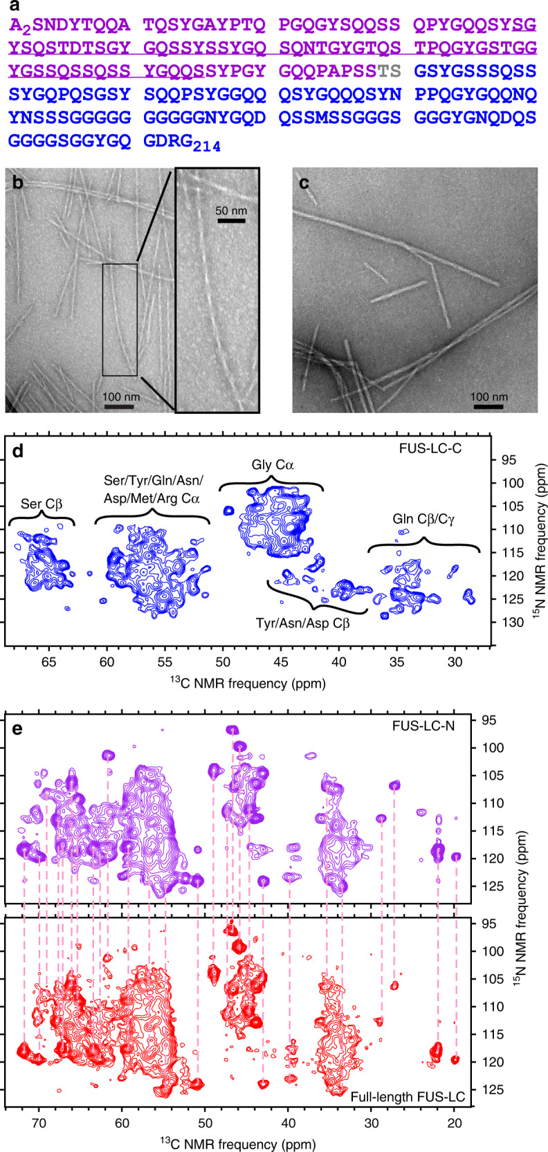
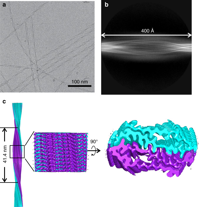
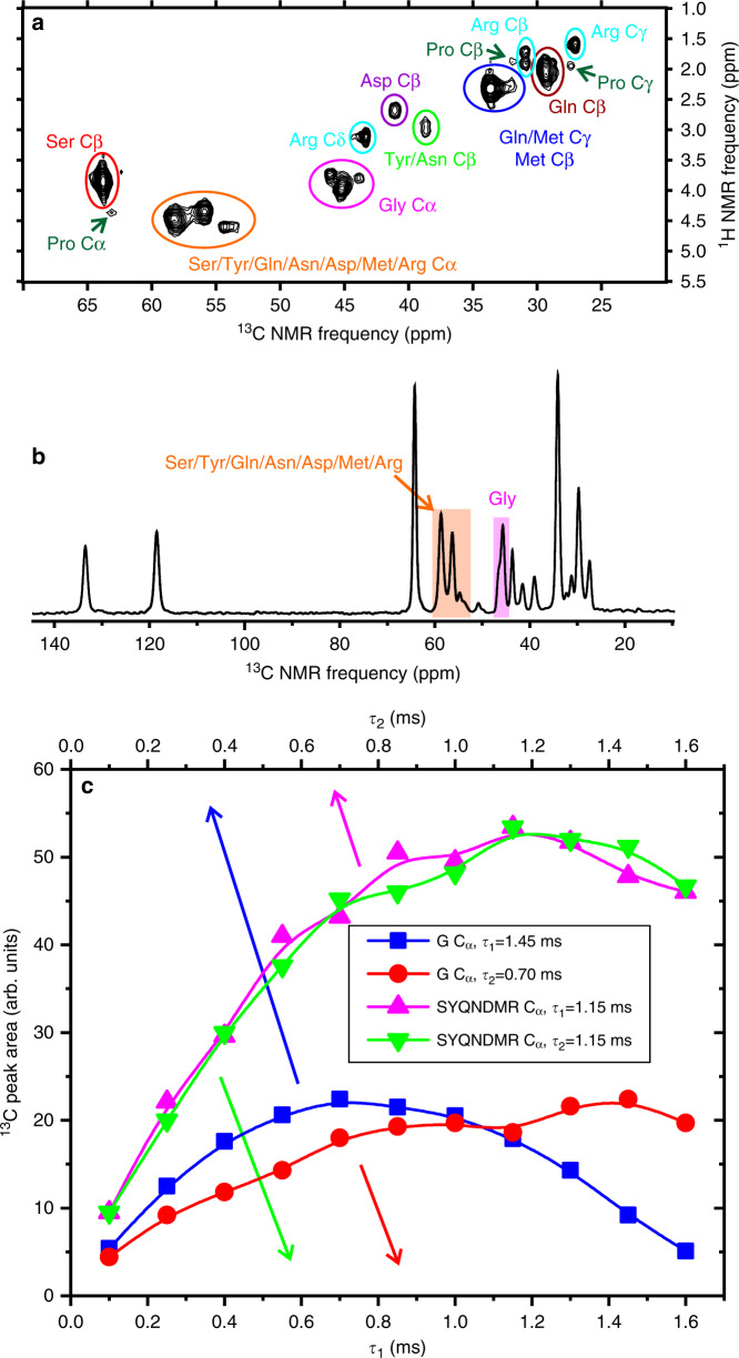
Protein domains without the usual distribution of amino acids, called low complexity (LC) domains, can be prone to self-assembly into amyloid-like fibrils. Self-assembly of LC domains that are nearly devoid of hydrophobic residues, such as the 214-residue LC domain of the RNA-binding protein FUS, is particularly intriguing from the biophysical perspective and is biomedically relevant due to its occurrence within neurons in amyotrophic lateral sclerosis, frontotemporal dementia, and other neurodegenerative diseases. We report a high-resolution molecular structural model for fibrils formed by the C-terminal half of the FUS LC domain (FUS-LC-C, residues 111-214), based on a density map with 2.62 Å resolution from cryo-electron microscopy (cryo-EM). In the FUS-LC-C fibril core, residues 112-150 adopt U-shaped conformations and form two subunits with in-register, parallel cross-β structures, arranged with quasi-21 symmetry. All-atom molecular dynamics simulations indicate that the FUS-LC-C fibril core is stabilized by a plethora of hydrogen bonds involving sidechains of Gln, Asn, Ser, and Tyr residues, both along and transverse to the fibril growth direction, including diverse sidechain-to-backbone, sidechain-to-sidechain, and sidechain-to-water interactions. Nuclear magnetic resonance measurements additionally show that portions of disordered residues 151-214 remain highly dynamic in FUS-LC-C fibrils and that fibrils formed by the N-terminal half of the FUS LC domain (FUS-LC-N, residues 2-108) have the same core structure as fibrils formed by the full-length LC domain. These results contribute to our understanding of the molecular structural basis for amyloid formation by FUS and by LC domains in general.


Conflict of interest statement

The authors have no competing interests.

Figures

Fig. 1
Fig. 1. Fibril formation by full-length FUS-LC and by its N-terminal and the C-terminal halves.
a Full-length FUS-LC sequence, residues 2–214. Segments in purple and blue lettering are FUS-LC-N and FUS-LC-C, respectively. The segment that forms the structurally ordered core of full-length FUS-LC fibrils is underlined. b Negative-stain TEM image of FUS-LC-C fibrils. Inset shows the rapidly twisting morphology. The same fibril morphology was observed in 53 images. c Negative-stain TEM image of FUS-LC-N fibrils. The same fibril morphologies were observed in 20 images. d 2D NCACX ssNMR spectra of FUS-LC-C fibrils, with residue-type assignments of cross-peak clusters. e 2D NCACX ssNMR spectra of uniformly 15N,13C-labeled FUS-LC-N fibrils (purple), and full-length FUS-LC fibrils (red). Vertical dashed lines, all with the same length, connect corresponding cross-peaks in the two spectra. All fibril samples were uniformly 15N,13C-labeled. Contour levels in all 2D spectra increase by successive factors of 1.3 and were set to show approximately the same number of levels below the maximum signals in all spectra.
Fig. 2
Fig. 2. Cryo-EM density map of FUS-LC-C fibrils.
a Representative cryo-EM image of FUS-LC-C fibrils. b Example of a 2D class average image, nearly spanning one crossover period. c 3D density map reconstruction from 275,520 particles. Nominal resolution is 2.63 Å. The density has a left-handed twist and is reconstructed with quasi-21 symmetry, defined by a helical rise of 2.44 Å and twist of 178.94°. Densities for the two cross-β subunits are colored cyan and purple.
Fig. 3
Fig. 3. Molecular structural model for the FUS-LC-C fibril core, consisting of residues 112–140.
a Cross-sectional view, with side-chain carbon atoms of Ser, Tyr, Gln, and Pro residues colored cyan, yellow, magenta, and green, respectively. b Side view, showing that the directions of backbone carbonyl groups in the molecular model align with backbone corrugations in the density map, consistent with the left-handed twist of the density map. c Expanded view of the interface between cross-β subunits. Pink asterisks indicate density attributable to ordered water molecules. d Cartoon representation of the molecular model, illustrating the 12° angle between β-strands of the two cross-β subunits (green and magenta) and 84° angle to the fibril growth direction (black arrow).
Fig. 4
Fig. 4. Detection of dynamically disordered segments by NMR.
a Aliphatic region of the 2D 1H-13C INEPT spectrum of uniformly 15N,13C-labeled FUS-LC-C fibrils. Residue-type assignments of cross-peaks are based on the known random-coil 1H and 13C chemical shifts of each residue type. b 1D 13C INEPT spectrum. Cα signals of Gly residues (pink) and other residues (orange) are highlighted. c Dependences of Cα peak areas in 1D INEPT 13C spectra on polarization transfer periods τ1 and τ2, for Gly residues (red and blue symbols, respectively) and other residues (green and pink symbols, respectively). Solid lines are guides to the eye.
Fig. 5
Fig. 5. Internal water and side-chain interactions in the FUS-LC-C fibril core, from all-atom molecular dynamics simulations in explicit solvent.
A cross-sectional slab that includes three repeats of the cryo-EM-based structure is shown after 400 ns of simulated dynamics, with position constraints on backbone Cα atoms. Side-chain carbon atoms of Ser, Tyr, Pro, and Gln or Asn residues are shown in cyan, yellow, green, and magenta, respectively. Oxygen atoms of internal and external water molecules are shown in red and orange, respectively. Significant features include (i) S116–S142 side-chain hydrogen-bonding chains along the fibril growth direction, (ii) intermolecular Gln–Gln polar zipper interactions along the growth direction, (iii) hydrogen bonds between Q126 side-chain amide and Q133 backbone carbonyl groups, (iv) hydrogen bonds between Q141 side-chain amide and G137 backbone carbonyl groups, (v) isolated internal water molecules, (vi) water within internal pores, lined with side chains of Ser, Tyr, and Gln residues, (vii) hydrogen bonds between Y149 side-chain hydroxyl and Y130 backbone carbonyl groups, and (viii) hydrogen bond networks involving side chains of Y136, Q147, and Q145.

References

    1. Banani SF, Lee HO, Hyman AA, Rosen MK. Biomolecular condensates: organizers of cellular biochemistry. Nat. Rev. Mol. Cell Biol. 2017;18:285–298. doi: 10.1038/nrm.2017.7. - DOI - PMC - PubMed
    1. Martin EW, Mittag T. Relationship of sequence and phase separation in protein low complexity regions. Biochemistry. 2018;57:2478–2487. doi: 10.1021/acs.biochem.8b00008. - DOI - PMC - PubMed
    1. Kato M, et al. Cell-free formation of RNA granules: Low complexity sequence domains form dynamic fibers within hydrogels. Cell. 2012;149:753–767. doi: 10.1016/j.cell.2012.04.017. - DOI - PMC - PubMed
    1. Lin Y, Protter DSW, Rosen MK, Parker R. Formation and maturation of phase-separated liquid droplets by RNA-binding proteins. Mol. Cell. 2015;60:208–219. doi: 10.1016/j.molcel.2015.08.018. - DOI - PMC - PubMed
    1. King OD, Gitler AD, Shorter J. The tip of the iceberg: RNA-binding proteins with prion-like domains in neurodegenerative disease. Brain Res. 2012;1462:61–80. doi: 10.1016/j.brainres.2012.01.016. - DOI - PMC - PubMed

Publication types