. 2020 Nov 12;10(1):19704.
doi: 10.1038/s41598-020-76898-2.

Functional impacts of the ubiquitin-proteasome system on DNA damage recognition in global genome nucleotide excision repair

Affiliations

Functional impacts of the ubiquitin-proteasome system on DNA damage recognition in global genome nucleotide excision repair

Wataru Sakai et al. Sci Rep. .

Abstract

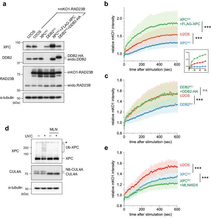
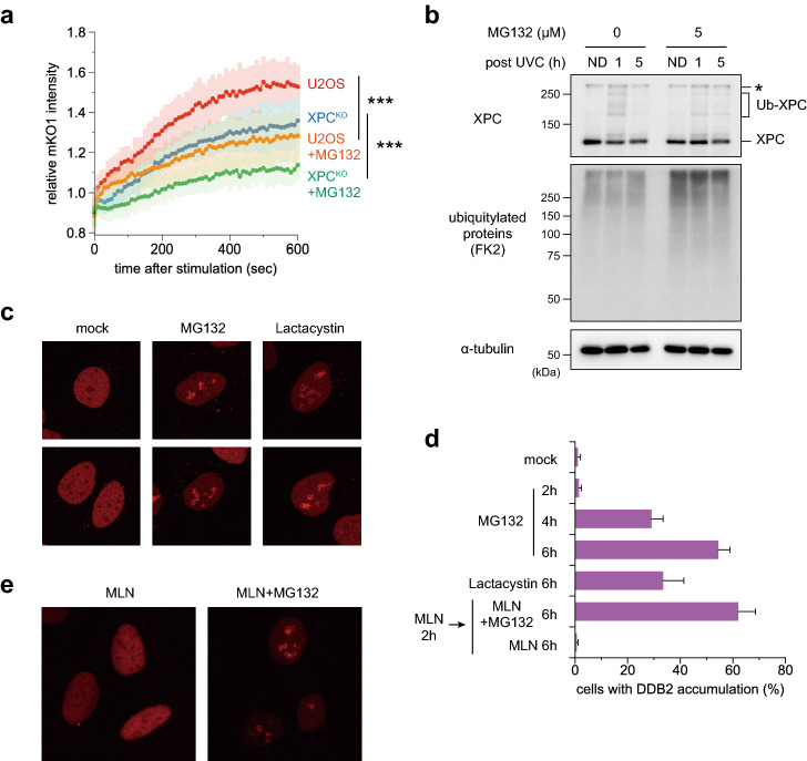
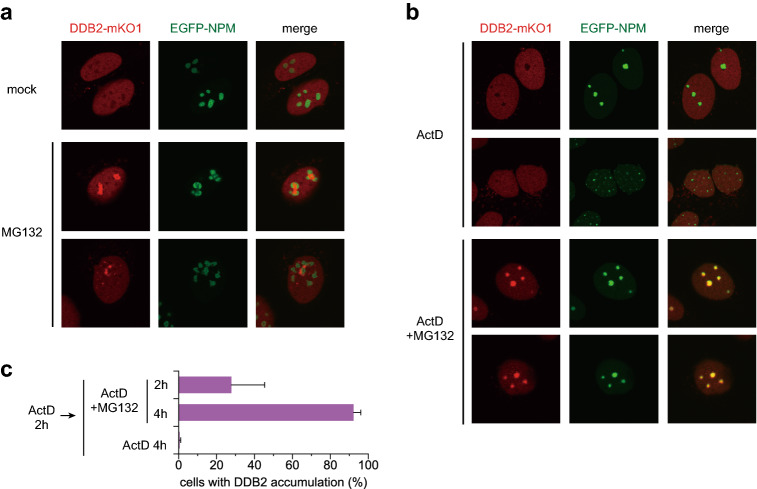
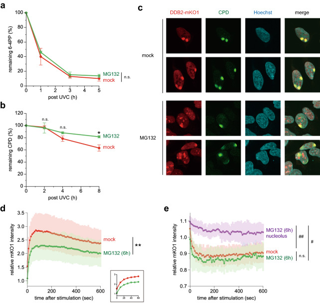
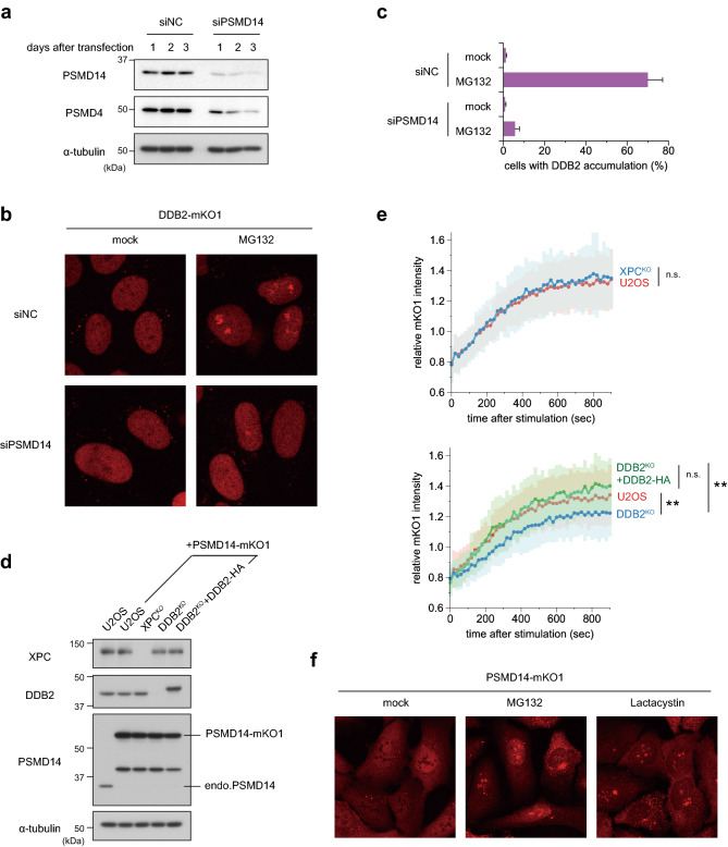
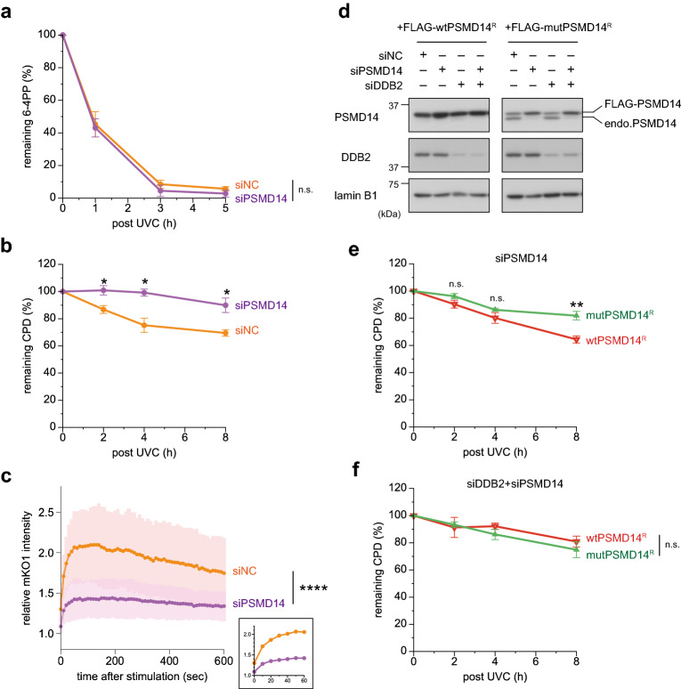
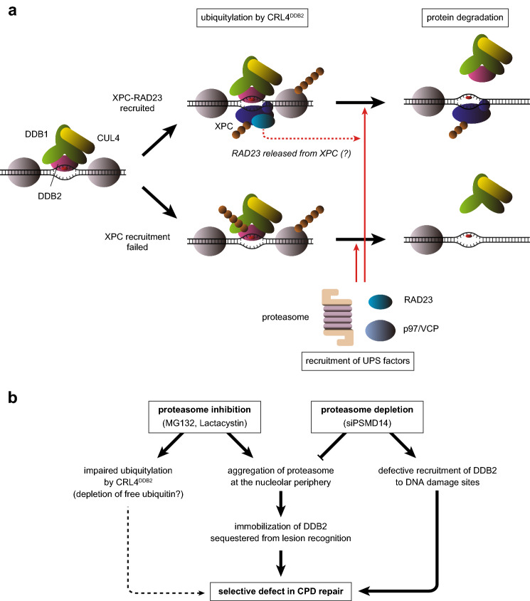
The ubiquitin-proteasome system (UPS) plays crucial roles in regulation of various biological processes, including DNA repair. In mammalian global genome nucleotide excision repair (GG-NER), activation of the DDB2-associated ubiquitin ligase upon UV-induced DNA damage is necessary for efficient recognition of lesions. To date, however, the precise roles of UPS in GG-NER remain incompletely understood. Here, we show that the proteasome subunit PSMD14 and the UPS shuttle factor RAD23B can be recruited to sites with UV-induced photolesions even in the absence of XPC, suggesting that proteolysis occurs at DNA damage sites. Unexpectedly, sustained inhibition of proteasome activity results in aggregation of PSMD14 (presumably with other proteasome components) at the periphery of nucleoli, by which DDB2 is immobilized and sequestered from its lesion recognition functions. Although depletion of PSMD14 alleviates such DDB2 immobilization induced by proteasome inhibitors, recruitment of DDB2 to DNA damage sites is then severely compromised in the absence of PSMD14. Because all of these proteasome dysfunctions selectively impair removal of cyclobutane pyrimidine dimers, but not (6-4) photoproducts, our results indicate that the functional integrity of the proteasome is essential for the DDB2-mediated lesion recognition sub-pathway, but not for GG-NER initiated through direct lesion recognition by XPC.


Conflict of interest statement

The authors declare no competing interests.

Figures

Figure 1
Figure 1
Real-time monitoring of recruitment of mKO1-RAD23B to local DNA damage. (a) Immunoblot analyses of endogenous and ectopically expressed XPC, DDB2, and RAD23B in the indicated cell lines. (b,c) Local DNA damage was induced in the indicated cell lines by three-photon absorption with an infrared femtosecond laser. Relative mKO1 intensities at damaged sites were quantitatively assessed over time. Inset in (b) shows enlargement of the graph in the early time range. The statistical significance assessed for the last time point is shown. *** P < 0.001. (d) Immunoblot analyses validating the effects of MLN4924. U2OS cells were pre-incubated for 2 h in the presence or absence of 1 µM MLN4924, and then irradiated with UVC at 10 J/m2 (or mock-irradiated) and further incubated for 1 h with or without MLN4924. Asterisk indicates non-specific reaction of the anti-XPC antibody. e The indicated cell lines were pre-treated for 2 h with or without 1 µM MLN4924, and recruitment of mKO1-RAD23B to local DNA damage was measured as in panels b and c. *** P < 0.001.
Figure 2
Figure 2
Inhibition of the proteasome affects dynamics and localization of GG-NER factors. (a) U2OS and XPCKO cells were pre-treated for 2 h with 5 µM MG132 or DMSO (solvent only). Recruitment of mKO1-RAD23B to local DNA damage was assessed as in Fig. 1. *** P < 0.001. (b) Immunoblot analyses of the effects of MG132. U2OS cells were pre-cultured for 2 h in the presence or absence of 5 µM MG132, irradiated with UVC at 10 J/m2, and further incubated for the indicated times with or without MG132. Asterisk indicates non-specific reaction of the anti-XPC antibody. ND: unirradiated control cells. (c) Representative images of the DDB2-mKO1 localization in the cells, which were mock-treated or treated for 6 h with 5 µM MG132 or 10 µM lactacystin. (d) Percentages of cells showing subnuclear DDB2-mKO1 accumulation after the indicated treatments. At least three independent experiments were carried out, and more than 300 cells in total were examined for each treatment. e Cells expressing DDB2-mKO1 were pre-incubated for 2 h in the presence of 1 µM MLN4924, and subsequently treated for 6 h with MLN4924 only (left) or with MLN4924 plus MG132 (right).
Figure 3
Figure 3
MG132-induced relocalization of DDB2 is related to nucleoli. (a) Cells expressing both DDB2-mKO1 and EGFP-NPM were mock-treated or incubated for 6 h in the presence of 5 µM MG132. Live-cell images were acquired with a confocal laser scanning microscope. (b) After 2-h pre-incubation with 40 nM ActD, cells were further treated for 4 h with ActD only or with ActD plus MG132. (c) Percentages of the cells showing DDB2-mKO1 accumulation were quantified as in Fig. 2d.
Figure 4
Figure 4
Inhibition of proteasome activity compromises GG-NER. (a,b) U2OS cells were pre-incubated for 2 h in the presence or absence of 5 µM MG132. The cells were then irradiated with UVC at 10 J/m2 (a) or 2 J/m2 (b), and further incubated for the indicated times with or without MG132. Amounts of 6-4PPs (a) and CPDs (b) remaining in genomic DNA were quantified by enzyme-linked immunosorbent assay with lesion-specific antibodies. * P < 0.05. (c) Cells expressing DDB2-mKO1 were pre-incubated for 6 h in the presence or absence of 5 µM MG132, and then irradiated with UVC at 100 J/m2 through isopore membrane filters. After a 10-min incubation with or without MG132, the cells were fixed and co-stained with anti-mKO2 and anti-CPD antibodies, and visualized with Alexa Fluor 594- and 488-conjugated secondary antibodies, respectively. Nuclear DNA was counter-stained with Hoechst 33342. (d) Cells expressing DDB2-mKO1 were pre-treated for 6 h with 5 µM MG132. The cells with nucleolar accumulation of DDB2 were chosen, and local UVC stimulation was applied to the areas outside the nucleoli. Recruitment of DDB2-mKO1 to the damaged sites was quantitatively assessed and compared with the data obtained with mock-treated cells. Inset shows enlargement of the graph in the early time range. The statistical significance assessed for the last time point is shown. ** P < 0.01. (e) Cells were treated as in (d), and fluorescence intensities of DDB2-mKO1 in the unstimulated areas were monitored. For MG132-treated cells, the areas with (purple line) or without (green line) the DDB2 accumulation were examined. # P < 1 × 10–8, ## P < 1 × 10–10.
Figure 5
Figure 5
MG132-induced DDB2 accumulation depends on the proteasome subunit PSMD14. (a) Immunoblot analyses validating depletion of the proteasome subunits. U2OS cells were transfected with negative control siRNA (siNC) or siRNA targeting PSMD14 (siPSMD14), and cell lysates were prepared after incubation for the indicated number of days. All subsequent experiments were performed on the second day of post-transfection. (b,c) Cells expressing DDB2-mKO1 were treated with siNC or siPSMD14, and then incubated for 6 h in the presence or absence of 5 µM MG132. Based on live-cell images acquired with a confocal laser scanning microscope (b), percentages of cells with subnuclear DDB2 accumulation were measured as in Fig. 2d. (d) Immunoblot analyses validating expression of the XPC, DDB2, and PSMD14 proteins in the indicated cell lines. (e) Recruitment of PSMD14-mKO1 to local DNA damage was assessed quantitatively in the indicated cell lines. ** P < 0.01. (f) Representative live-cell images of PSMD14-mKO1 localization in cells that were mock-treated or treated for 6 h in the presence of 5 µM MG132 or 10 µM lactacystin.
Figure 6
Figure 6
Presence of a functional proteasome is required for efficient CPD repair. (a,b) U2OS cells treated with siNC or siPSMD14 were used to assess repair kinetics of 6-4PPs (a) and CPDs (b) as in Fig. 4. * P < 0.05. (c) Cells expressing DDB2-mKO1 were treated with siNC or siPSMD14, and recruitment of DDB2-mKO1 to local DNA damage was quantitatively assessed. The statistical significance assessed for the last time point is shown. **** P < 1 × 10–4. (d) Immunoblot analyses validating the effects of siRNAs and the resistance of the ectopically-expressed PSMD14R proteins to the siPSMD14. (e,f) Cells expressing wtPSMD14R or mutPSMD14R were treated with siPSMD14 only (e) or siDDB2 plus siPSMD14 (f). After UVC irradiation at 2 J/m2, repair kinetics of CPDs were assessed. ** P < 0.01.
Figure 7
Figure 7
Schematic representation of functional impacts of UPS on GG-NER. (a) Model for roles of UPS factors in the GG-NER process. The CRL4DDB2 E3 ligase bound to a DNA lesion ubiquitylates DDB2, XPC, and other proteins nearby (such as histones). Successful recruitment of XPC-RAD23 competitively suppresses ubiquitylation and degradation of DDB2. UPS factors such as the proteasome, RAD23, and p97/VCP are then recruited to degrade poly-ubiquitylated substrates. (b) Impacts of proteasome dysfunctions revealed by this study.

Similar articles

Cited by

References

    1. Marteijn JA, Lans H, Vermeulen W, Hoeijmakers JH. Understanding nucleotide excision repair and its roles in cancer and ageing. Nat. Rev. Mol. Cell Biol. 2014;15:465–481. doi: 10.1038/nrm3822. - DOI - PubMed
    1. Schärer OD. Nucleotide excision repair in eukaryotes. Cold Spring Harb. Perspect. Biol. 2013;5:a012609. doi: 10.1101/cshperspect.a012609. - DOI - PMC - PubMed
    1. Gillet LCJ, Schärer OD. Molecular mechanisms of mammalian global genome nucleotide excision repair. Chem. Rev. 2006;106:253–276. doi: 10.1021/cr040483f. - DOI - PubMed
    1. DiGiovanna JJ, Kraemer KH. Shining a light on xeroderma pigmentosum. J. Invest. Dermatol. 2012;132:785–796. doi: 10.1038/jid.2011.426. - DOI - PMC - PubMed
    1. Nishigori C, Sugasawa K, editors. DNA Repair Disorders. New York: Springer Nature; 2019.

Publication types