Review
. 2021 Jan;22(1):38-53.
doi: 10.1038/s41583-020-00396-7. Epub 2020 Nov 12.

Encouraging an excitable brain state: mechanisms of brain repair in stroke

Affiliations
Review

Encouraging an excitable brain state: mechanisms of brain repair in stroke

Mary T Joy et al. Nat Rev Neurosci. 2021 Jan.

Abstract

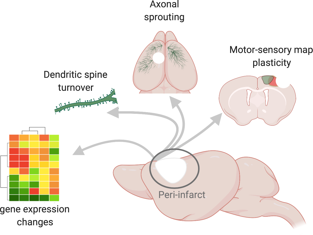
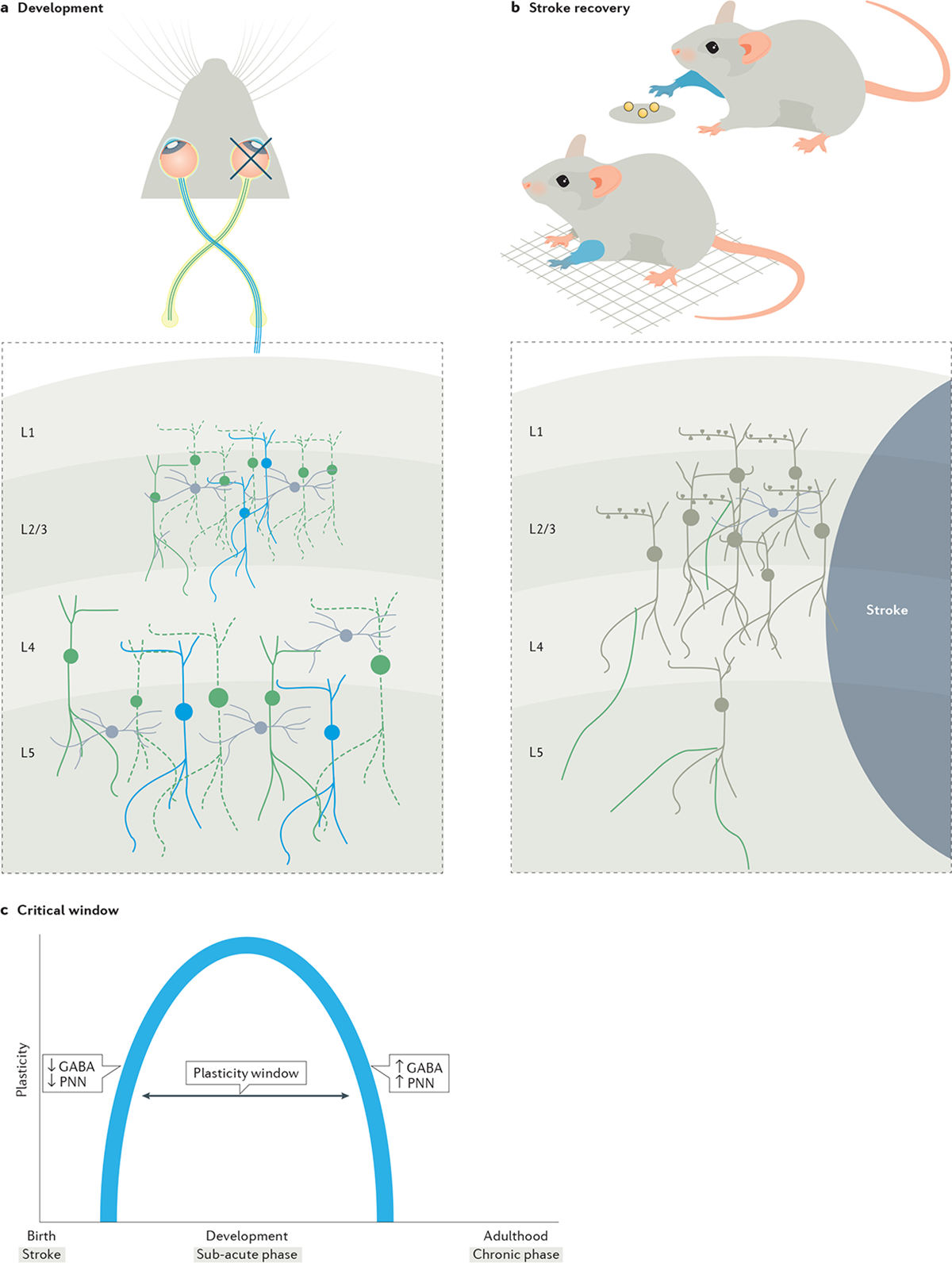
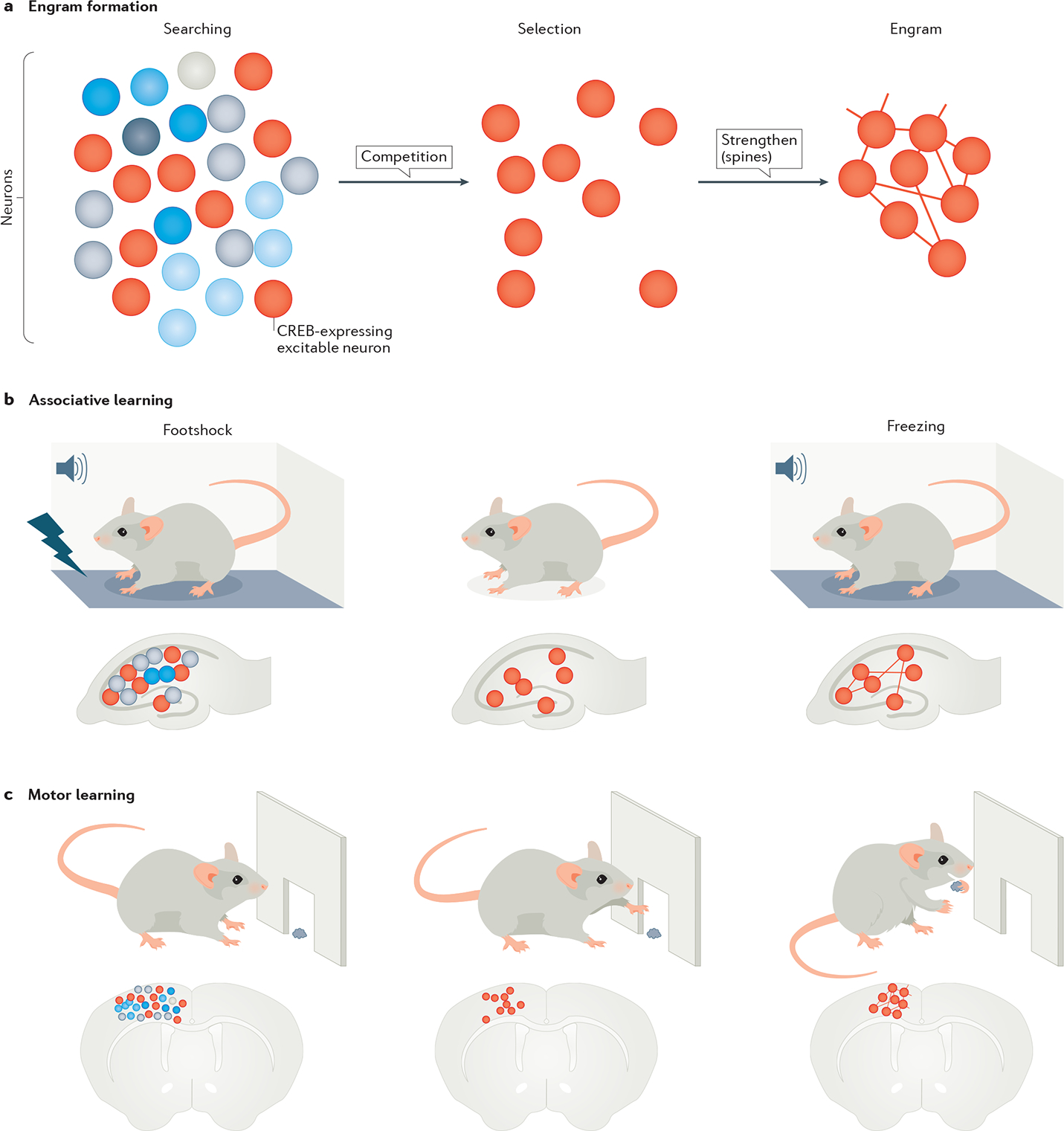
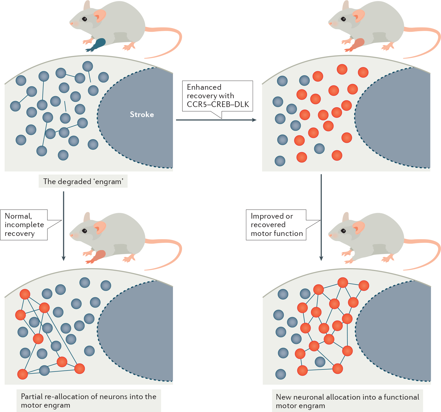
Stroke induces a plastic state in the brain. This period of enhanced plasticity leads to the sprouting of new axons, the formation of new synapses and the remapping of sensory-motor functions, and is associated with motor recovery. This is a remarkable process in the adult brain, which is normally constrained in its levels of neuronal plasticity and connectional change. Recent evidence indicates that these changes are driven by molecular systems that underlie learning and memory, such as changes in cellular excitability during memory formation. This Review examines circuit changes after stroke, the shared mechanisms between memory formation and brain repair, the changes in neuronal excitability that underlie stroke recovery, and the molecular and pharmacological interventions that follow from these findings to promote motor recovery in animal models. From these findings, a framework emerges for understanding recovery after stroke, central to which is the concept of neuronal allocation to damaged circuits. The translation of the concepts discussed here to recovery in humans is underway in clinical trials for stroke recovery drugs.


Figures

Fig. 1 |
Fig. 1 |. Endogenous plasticity in sub-acute stroke.
Sagittal view of a mouse brain (centre) in which the site of a stroke is depicted by the white area and the peri-infarct region is demarcated by a circle. The arrows point to molecular and cellular hallmarks of the peri-infarct region, including changes in gene expression. Here, these changes are denoted as a heatmap schematic in which the magnitudes of the changes in gene expression are visualized as differently coloured modules. New molecular programs support structural connectivity such as strengthening of connections through the growth and turnover of new spines, which are post-synaptic protrusions on dendrites, and the growth of new axons, depicted as blue fibres that sprout from the peri-infarct cortex and travel to intact motor, somatosensory and contralateral cortices. Changes in structural connectivity may underlie re-organization of functional motor (blue) and somatosensory (green) limb representations that re-appear at new locations several weeks after a stroke.
Fig. 2 |
Fig. 2 |. Parallels between windows of plasticity in development and stroke.
The critical period in development and the sensitive period in stroke share molecular and connectional principles. a | Visual experience during the critical period shapes neuronal connectivity. Temporary deprivation of visual experience, only during the critical period, by unilateral eye closure (depicted by the cross on one eye), can cause a decrease in the responsiveness of neurons (dotted neurons in green) to a visual stimulus across different layers of the visual cortex pertaining to the closed eye. Here, during this window, neural connectivity is amenable to plastic changes and shaped by visual experience. b | After a stroke, a similar window of plasticity exists. Stroke impairs motor function such as in the ability of a stroke-induced mouse to locomote on a grid or in skilled reaching of a food pellet with the impaired limb (depicted as blue forelimbs). The sub-acute phase of stroke is marked by a sensitive period similar to the critical period in development in which turnover of dendritic spines in the peri-infarct cortex and the sprouting of new axons (green axons on neurons) provide a neural substrate for functional reorganization of limb representations (not shown). These endogenous mechanisms are associated with spontaneous incomplete recovery of motor function, in which the impaired limb gains limited ability or compensates to perform a motor task. c | The opening and closing of the critical window during development and the sensitive period in stroke as it progresses from acute to chronic are regulated by similar molecular mechanisms, such as changes in excitatory to inhibitory GABA signalling and the maturation of peri-neuronal nets (PNNs).
Fig. 3 |
Fig. 3 |. Storage of memory in co-active neuronal networks, or engrams.
The figure shows the stages of engram formation that store a fear memory or motor memory. a | Engram formation begins with active searching for neurons that are co-active during a behavioural event. From this pool of neurons with similar spatiotemporal activation patterns, neurons with higher cAMP-binding response element (CREB) expression (red circles) are selected to form an engram. The connections within an engram are further strengthened through their dendritic spines that carry post-synaptic information. b | This part shows a collection of neurons in the hippocampus, with those showing CREB expression (red circles) storing a fear memory by associating foot shock with a tone. The fear memory can be recalled by reproducing the tone in the absence of shock and leads to freezing behaviour. The process of recall involves reactivating the memory engram associated with the foot shock. c | This part shows a similar engram in the motor cortex, which stores a motor skill as the motor task is being learnt. The learned function is allocated to a sparse and specific set of neurons initially selected from a large network.
Fig. 4 |
Fig. 4 |. Recovery engram in stroke.
A conceptual model of the engram in stroke recovery. Top left panel: stroke induces a loss of neural connectivity, disrupting the engram that encodes a motor behaviour and, hence, causing a motor impairment. Note that the level of functional impairment is depicted by the colour of the mouse forelimb, ranging from blue (no function) to shades of red (partial recovery) and grey (normal movement). Bottom left panel: endogenous plasticity during the critical period provides a new neural substrate where lost function is partially allocated to a sub-set of neurons (red circles) in a motor circuit. Inefficient allocation leads to spontaneous partial recovery or compensation of motor function. Right panels: increasing the excitability threshold of neurons in peri-infarct by increasing cAMP-binding response element (CREB) expression captures excitable and efficient neurons to allocate into a functional motor circuit for complete motor recovery. CREB–C-C chemokine receptor 5 (CCR5)–dual-leucine zipper kinase (DLK) signalling, which can mediate allocation, may be a target in developing drugs for stroke recovery.

Similar articles

Cited by

References

    1. Benjamin EJ et al. Heart Disease and Stroke Statistics-2019 Update: A Report From the American Heart Association. Circulation 139, e56–e528 (2019). - PubMed
    1. Faul M, Xu L, Wald MM & Coronado VG Traumatic brain injury in the United States: emergency department visits, hospitalizations, and deaths. Atlanta (GA): Centers for Disease Control and Prevention. Natl. Cent. Inj. Prev. Control 2, (2010).
    1. Song SS Advanced imaging in acute ischemic stroke. Semin. Neurol. 33, 436–440 (2013). - PubMed
    1. Fisher M & Albers GW Advanced imaging to extend the therapeutic time window of acute ischemic stroke. Ann. Neurol. 73: 4–9 (2013). - PubMed
    1. Sandhu GS & Sunshine JL Advanced neuroimaging to guide acute stroke therapy. Curr. Cardiol. Rep. 14, 741–753 (2012). - PubMed

Publication types