

Factors associated with extremely poor visual outcomes in patients with central retinal vein occlusion
- PMID: 33184484
- PMCID: PMC7665063
- DOI: 10.1038/s41598-020-76840-6
Factors associated with extremely poor visual outcomes in patients with central retinal vein occlusion
Abstract
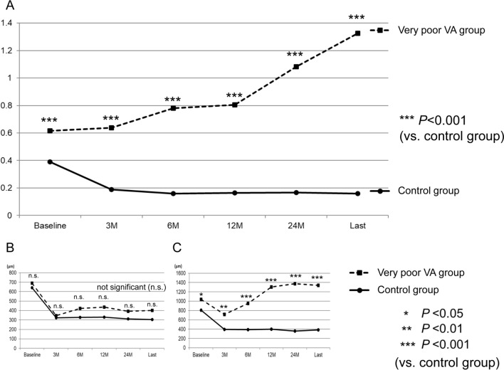
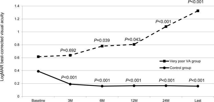
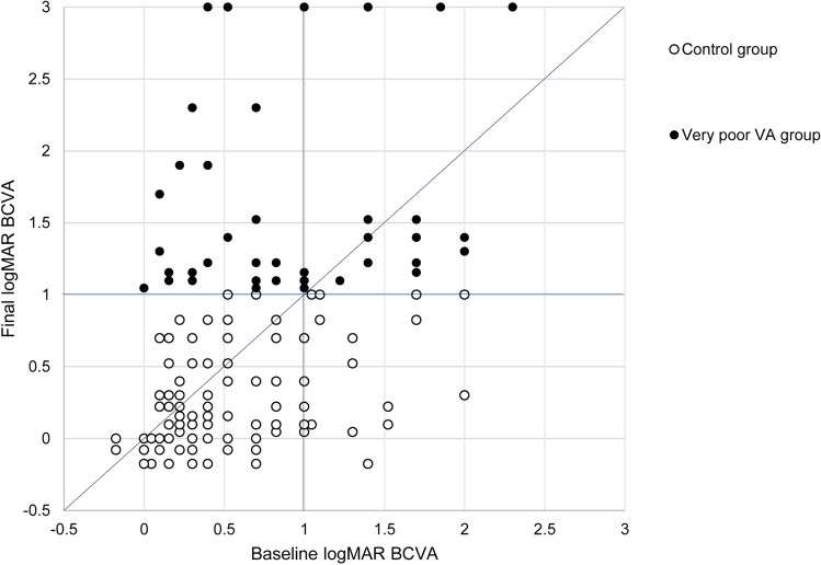
Here, we examined prognostic factors for extremely poor visual outcomes in patients with central retinal vein occlusion (CRVO) in actual practices. We included 150 consecutive eyes with treatment-naïve acute CRVO from four different facilities and observed them for over 24 months. Macular edema (ME) was treated with one or three monthly anti-vascular endothelial growth factor injections (1 or 3 + pro re nata). According to the final Snellen visual acuity (VA), we divided the patients into very poor VA (< 20/200) and control (≥ 20/200) groups and examined risk factors for poor final visual outcomes. The baseline Snellen VA was hand motion to 20/13. The mean number of anti-VEGF injections for ME was 5.3 ± 3.7 during the follow-up period. In total, 49 (32.7%) patients exhibited a very poor final VA; this group comprised significantly older patients with a significantly poorer baseline VA (P < 0.01 for both) than the control group. Comorbid internal carotid artery disease and diabetic retinopathy were significantly associated with a poor final VA. In actual clinical practice, visual outcomes may be extremely poor despite ME treatment in certain patients with CRVO, with advanced age, poor baseline VA, and comorbid internal carotid artery disease and diabetic retinopathy being significant risk factors.
Conflict of interest statement
The authors declare no competing interests.
Figures
Similar articles
-
Association of Disorganization of Retinal Inner Layers With Visual Acuity Response to Anti-Vascular Endothelial Growth Factor Therapy for Macular Edema Secondary to Retinal Vein Occlusion.JAMA Ophthalmol. 2019 Jan 1;137(1):38-46. doi: 10.1001/jamaophthalmol.2018.4484. JAMA Ophthalmol. 2019. PMID: 30286219 Free PMC article.
-
Prospective study of intravitreal ranibizumab as a treatment for decreased visual acuity secondary to central retinal vein occlusion.Am J Ophthalmol. 2009 Feb;147(2):298-306. doi: 10.1016/j.ajo.2008.08.016. Epub 2008 Oct 17. Am J Ophthalmol. 2009. PMID: 18929354 Clinical Trial.
-
Impact of initial visual acuity on anti-VEGF treatment outcomes in patients with macular oedema secondary to retinal vein occlusions in routine clinical practice.Br J Ophthalmol. 2017 May;101(5):574-579. doi: 10.1136/bjophthalmol-2016-308727. Epub 2016 Aug 8. Br J Ophthalmol. 2017. PMID: 27503394
-
Early treatment of severe cystoid macular edema in central retinal vein occlusion with posterior sub-tenon triamcinolone acetonide.Retina. 2007 Feb;27(2):180-9. doi: 10.1097/01.iae.0000237584.56552.1c. Retina. 2007. PMID: 17290200
-
Comparison between Ozurdex and intravitreal anti-vascular endothelial growth factor treatment for retinal vein occlusion-related macular edema: A systematic review and meta-analysis of randomized controlled trials.Indian J Ophthalmol. 2019 Nov;67(11):1800-1809. doi: 10.4103/ijo.IJO_382_19. Indian J Ophthalmol. 2019. PMID: 31638037 Free PMC article.
Cited by
-
A retrospective study assessing the factors associated with visual outcome in retinal vein occlusion patients after anti-VEGF therapy.PeerJ. 2021 Dec 6;9:e12599. doi: 10.7717/peerj.12599. eCollection 2021. PeerJ. 2021. PMID: 34963823 Free PMC article.
-
Nomogram Prediction Model for Diabetic Retinopathy Development in Type 2 Diabetes Mellitus Patients: A Retrospective Cohort Study.J Diabetes Res. 2021 Sep 14;2021:3825155. doi: 10.1155/2021/3825155. eCollection 2021. J Diabetes Res. 2021. PMID: 34595241 Free PMC article.
-
Foveal Thickness Fluctuations in Anti-VEGF Treatment for Central Retinal Vein Occlusion.Ophthalmol Sci. 2023 Oct 29;4(2):100418. doi: 10.1016/j.xops.2023.100418. eCollection 2024 Mar-Apr. Ophthalmol Sci. 2023. PMID: 38146527 Free PMC article.
-
Prognosis and factors related to anti-VEGF therapy in patients with retinal vein occlusion and concomitant carotid artery disease.Sci Rep. 2024 Oct 20;14(1):24634. doi: 10.1038/s41598-024-75604-w. Sci Rep. 2024. PMID: 39428411 Free PMC article.
-
Background Factors Affecting Visual Acuity at Initial Visit in Eyes with Central Retinal Vein Occlusion: Multicenter Study in Japan.J Clin Med. 2021 Nov 29;10(23):5619. doi: 10.3390/jcm10235619. J Clin Med. 2021. PMID: 34884321 Free PMC article.
References
-
- Mruthyunjaya P, Fekrat S, et al. Central retinal vein occlusion. In: Ryan SJ, et al., editors. retina. 4. Amsterdam: Elsevier Mosby; 2006. pp. 1339–1348.
Publication types
MeSH terms
Substances
LinkOut - more resources
Full Text Sources
Medical
