. 2020 Nov 12;10(1):19630.
doi: 10.1038/s41598-020-76781-0.

The emerging role of neutrophil extracellular traps in severe acute respiratory syndrome coronavirus 2 (COVID-19)

Affiliations

The emerging role of neutrophil extracellular traps in severe acute respiratory syndrome coronavirus 2 (COVID-19)

Angélica Arcanjo et al. Sci Rep. .

Abstract

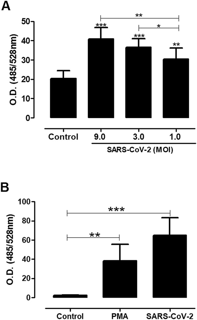
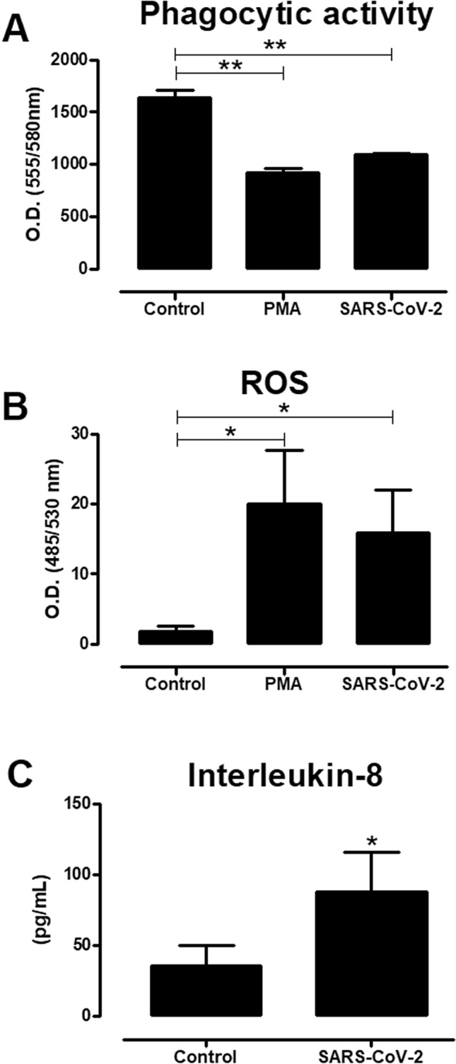
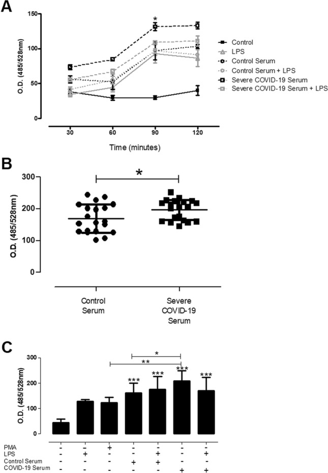
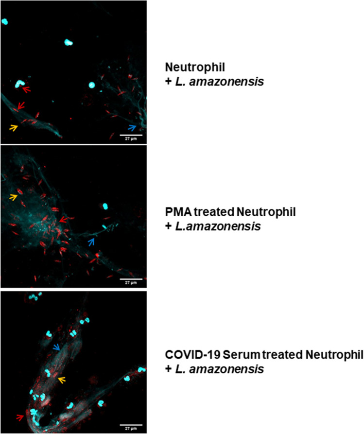
The novel coronavirus SARS-CoV-2 causes COVID-19, a highly pathogenic viral infection threatening millions. The majority of the individuals infected are asymptomatic or mildly symptomatic showing typical clinical signs of common cold. However, approximately 20% of the patients can progress to acute respiratory distress syndrome (ARDS), evolving to death in about 5% of cases. Recently, angiotensin-converting enzyme 2 (ACE2) has been shown to be a functional receptor for virus entry into host target cells. The upregulation of ACE2 in patients with comorbidities may represent a propensity for increased viral load and spreading of infection to extrapulmonary tissues. This systemic infection is associated with higher neutrophil to lymphocyte ratio in infected tissues and high levels of pro-inflammatory cytokines leading to an extensive microthrombus formation with multiorgan failure. Herein we investigated whether SARS-CoV-2 can stimulate extracellular neutrophils traps (NETs) in a process called NETosis. We demonstrated for the first time that SARS-CoV-2 in fact is able to activate NETosis in human neutrophils. Our findings indicated that this process is associated with increased levels of intracellular Reactive Oxygen Species (ROS) in neutrophils. The ROS-NET pathway plays a role in thrombosis formation and our study suggest the importance of this target for therapy approaches against disease.


Conflict of interest statement

The authors declare no competing interests.

Figures

Figure 1
Figure 1
Neutrophils release extracellular DNA in response to coronavirus 2 (SARS-CoV-2). Neutrophils (5 × 104/well) were stimulated or not with SARS-CoV-2 at different MOI ratios (A) or fixed MOI of 9.0 (B). The cells were incubated with virus or PMA (positive control) and after 90 min the supernatants were collected and the extracellular DNA release quantified by the Quant-iT PicoGreen dsDNA method, specific reagent for double-stranded DNA labeling in optical density assay (528 nm). The baseline control autofluorescence for the excitation/emission wavelengths (485/538 nm) is 6.30 ± 2.14. The data are representative of three independent experiments. The results were analyzed using One-way ANOVA followed by Tukey’s. Differences between groups are significant *p ≤ 0.05, **p ≤ 0.0025, ***p ≤ 0.0001.
Figure 2
Figure 2
Active coronavirus 2 (SARS-CoV-2) potentiates the activation of neutrophil extracellular traps (NETs) formation. Neutrophils (5 × 104/well) were stimulated alternatively with live or heat-inactivated SARS-CoV-2 (MOI of 9.0), and PMA (positive control). After 90 min of incubation, the coverslips were probed using anti-rabbit myeloperoxidase (Abcam 9535) and HOECHST (DNA marker). Arrows indicate the formation of structural NETs, showing the DNA (blue) and myeloperoxidase (red) markers. The images were made using an Olympus BX51 fluorescence microscope.
Figure 3
Figure 3
Neutrophils activate reactive oxygen species (ROS) and IL-8 secretion in response to coronavirus 2 (SARS-CoV-2). Neutrophils (5 × 104/well) were stimulated or not with SARS-CoV-2 virus (MOI of 9.0) or PMA (positive control), and after 90 min of incubation the quantification of reactive oxygen species (ROS) was determined (A). Intracellular ROS was quantified using H2DCFDA probe (see “Materials and methods”), and the values expressed refer to the quantification of florescence incorporated by the probe (530 nm). (B) The phagocytic activity was analyzed by uptake of Dextran beads conjugated to tetramethyl rhodamine (2,000,000 MW) added to the cells (200 µg/mL) in the presence of the stimuli. The values, expressed in optical density (580 nm), refer to the incorporated florescence after 90 min of incubation. (C) The quantification of interleukin-8 (IL-8) production from neutrophils stimulated with SARS-CoV-2 virus (MOI of 9.0) was measured from culture supernatants collected after a 90-min incubation according to the recommendations of the manufacturer Quantikine Elisa/R&D System (see “Materials and methods”). The baseline control autofluorescence for the excitation/emission wavelengths 485/530 nm is 3.43 ± 1.79; and 555/580 nm is 348.85 ± 112.03. The results were analyzed using paired t-test analysis and One-way ANOVA followed by Tukey’s. Differences between groups are significant *p ≤ 0.05, **p ≤ 0.0025.
Figure 4
Figure 4
Neutrophils release extracellular DNA in response to sera from patients with severe acute respiratory syndrome coronavirus 2 (SARS-CoV-2) infection. Neutrophils (5 × 104/well) were stimulated or not with LPS (10 µg/mL) in the presence of 10% sera from normal donors or severe patients in the acute phase of infection, after the different stimulation times indicated in the kinetics (A), or at a time point of 90 min (B,C). The supernatants were then collected and NETs were quantified by the Quant-iT PicoGreen dsDNA method, using specific reagent for double-stranded DNA detection by optical density (528 nm). The data are representative of three independent experiments. (A) Sera from two donors (healthy control and severe COVID-19 patient) were used; (B,C) Sera from a cohort of 40 donors (20 healthy controls and severe 20 severe COVID-19 patients in the acute phase of infection) were used. The results were analyzed using paired t-test analysis and One-way ANOVA followed by Tukey’s. Differences between groups are significant *p ≤ 0.05, **p ≤ 0.0025.
Figure 5
Figure 5
Neutrophil extracellular trap activated in COVID-19 are structurally and functionally active with ability to mediate pathogen entrapment. Neutrophils (2 × 105) were incubated with 100 nM PMA (Sigma) or 10% serum from severe COVID-19 patients in the acute phase of infection for 90 min. Afterwards, Leishmania amazonensis promastigotes were added to the culture (10 parasites per neutrophil) and incubated for 2 h at 35 °C for immunocytochemistry analysis. The coverslips were probed using anti-rabbit myeloperoxidase (Abcam 9535) and HOECHST (DNA marker). Arrows indicate the formation of structural NETs, showing the DNA (blue) and myeloperoxidase (red) markers. Yellow arrows indicate the presence of entrapped Leishmania parasites. The images were made using an Olympus BX51 fluorescence microscope.

References

    1. Rothan HA, et al. The epidemiology and pathogenesis of coronavirus disease (COVID-19) outbreak. J. Autoimmun. 2020;109:102433. doi: 10.1016/j.jaut.2020.102433. - DOI - PMC - PubMed
    1. Jin Y, et al. Virology, epidemiology, pathogenesis, and control of COVID-19. Viruses. 2020;12(4):372. doi: 10.3390/v12040372. - DOI - PMC - PubMed
    1. Ye Q, et al. The pathogenesis and treatment of the `Cytokine Storm' in COVID-19. J. Infect. 2020;80(6):607–613. doi: 10.1016/j.jinf.2020.03.037. - DOI - PMC - PubMed
    1. Bourgonje AR, et al. Angiotensin-converting enzyme-2 (ACE2), SARS-CoV-2 and pathophysiology of coronavirus disease 2019 (COVID-19) J. Pathol. 2020;251:228–248. doi: 10.1002/path.5471. - DOI - PMC - PubMed
    1. Raj K, et al. SARS-CoV-2 shedding from asymptomatic patients: Contribution of potential extrapulmonary tissue reservoirs. Am. J. Trop. Med. Hyg. 2020;103:18–21. doi: 10.4269/ajtmh.20-0279. - DOI - PMC - PubMed

Publication types

Substances