. 2020 Jun 24;13(5):447-461.
doi: 10.1007/s12195-020-00626-z. eCollection 2020 Oct.

Re-engineering Antimicrobial Peptides into Oncolytics Targeting Drug-Resistant Ovarian Cancers

Affiliations

Re-engineering Antimicrobial Peptides into Oncolytics Targeting Drug-Resistant Ovarian Cancers

Matthew R Aronson et al. Cell Mol Bioeng. .

Abstract

Introduction: Bacteria and cancer cells share a common trait-both possess an electronegative surface that distinguishes them from healthy mammalian counterparts. This opens opportunities to repurpose antimicrobial peptides (AMPs), which are cationic amphiphiles that kill bacteria by disrupting their anionic cell envelope, into anticancer peptides (ACPs). To test this assertion, we investigate the mechanisms by which a pathogen-specific AMP, originally designed to kill bacterial Tuberculosis, potentiates the lytic destruction of drug-resistant cancers and synergistically enhances chemotherapeutic potency.

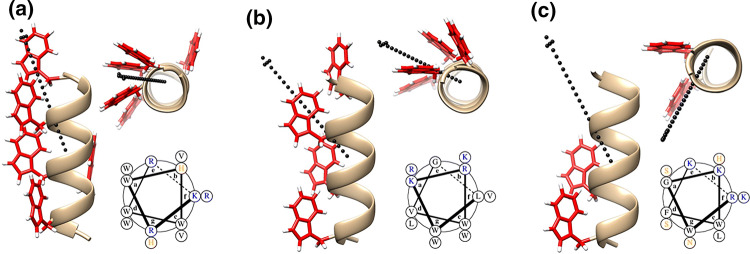
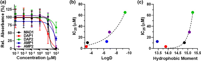
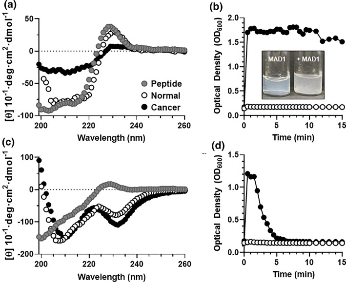
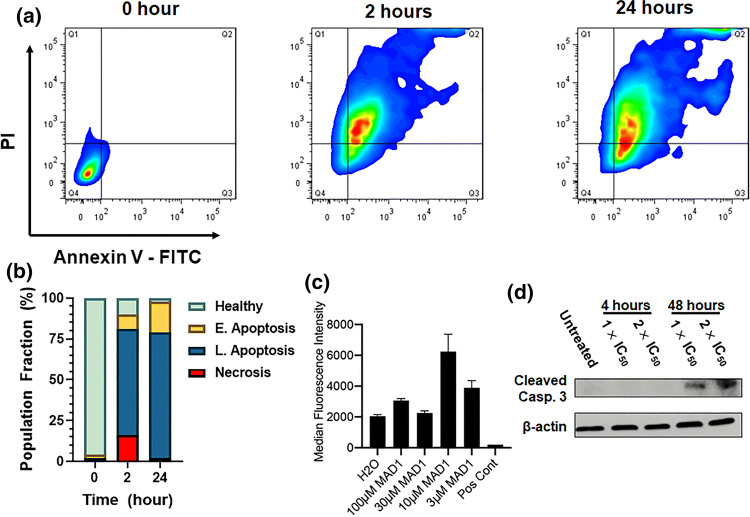
Materials and methods: De novo peptide design, paired with cellular assays, elucidate structure-activity relationships (SAR) important to ACP potency and specificity. Using the sequence MAD1, microscopy, spectrophotometry and flow cytometry identify the peptide's anticancer mechanisms, while parallel combinatorial screens define chemotherapeutic synergy in drug-resistant cell lines and patient derived ex vivo tumors.

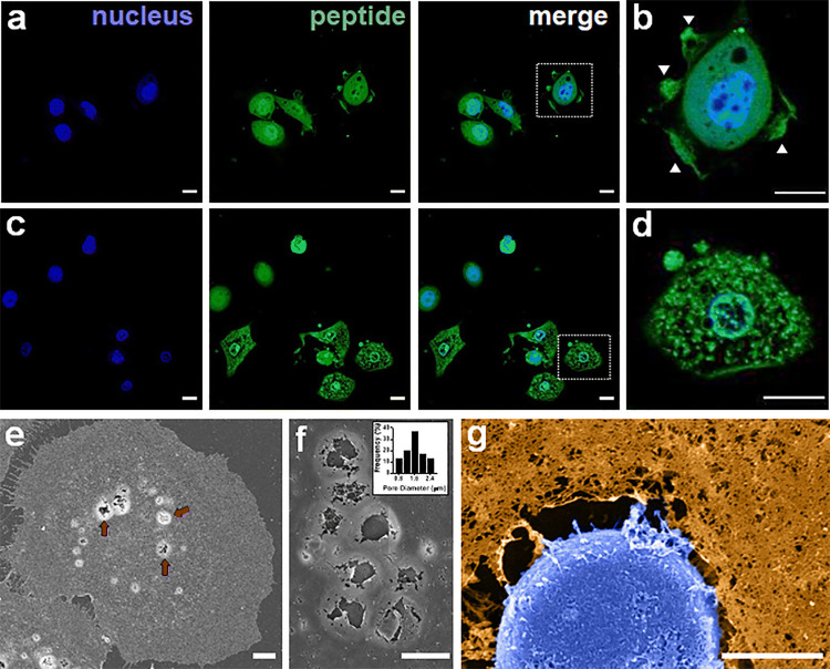
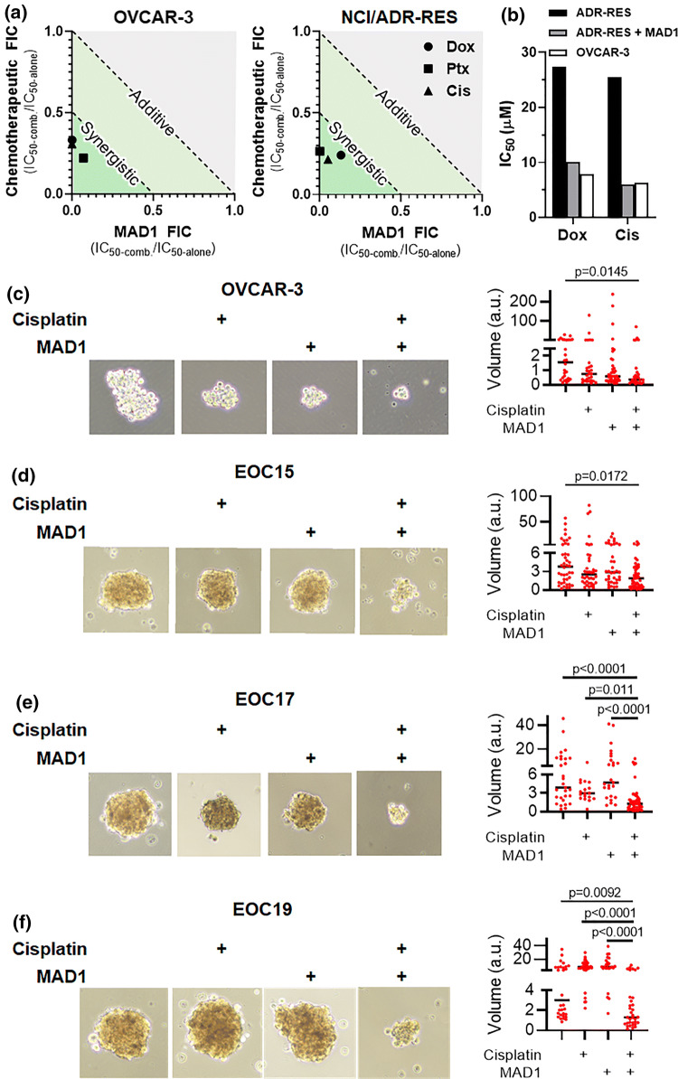
Results: SAR investigations reveal spatial sequestration of amphiphilic regions increases ACP potency, but at the cost of specificity. Selecting MAD1 as a lead sequence, mechanistic studies identify that the peptide forms pore-like supramolecular assemblies within the plasma and nuclear membranes of cancer cells to potentiate death through lytic and apoptotic mechanisms. This diverse activity enables MAD1 to synergize broadly with chemotherapeutics, displaying remarkable combinatorial efficacy against drug-resistant ovarian carcinoma cells and patient-derived tumor spheroids.

Conclusions: We show that cancer-specific ACPs can be rationally engineered using nature's AMP toolbox as templates. Selecting the antimicrobial peptide MAD1, we demonstrate the potential of this strategy to open a wealth of synthetic biotherapies that offer new, combinatorial opportunities against drug resistant tumors.

Keywords: Anticancer peptides; Combinatorial therapy; De novo design; Nanostructures; Supramolecular assembly.


Figures

Figure 1
Figure 1
Rational design of de novo anticancer peptides. Minimized model of (a) MAD1, (b) DAP1, (c) DAP2 peptide helices. For all panels, Left: helical profile; Top right: axial view (red = tryptophan residues). Vector of the hydrophobic moment shown as dotted black arrow. Bottom right: helical wheel (black = hydrophobic, blue = basic, orange = polar residues).
Figure 2
Figure 2
ACP cytotoxicity and structure-activity relationships. (a) Cytotoxicity curves for MAD1, DAP1, DAP2, AMP1, and AMP2 peptides against OVCAR-3 ovarian carcinoma cells, shown as percent relative MTT absorbance. Curves for all four cancer cell lines tested are shown in Supplementary Fig. 6. Relative change in OVCAR-3 IC25 value as a function of (b) sequence hydrophobicity (logD) and (c) helix facial amphiphilicity (hydrophobic moment). Exponential fit (dashed line, GraphPad Prism) included to guide the eye.
Figure 3
Figure 3
Tumor membrane-templated ACP assembly. (a) Circular dichroism spectra of the MAD1 peptide in aqueous solution (●), or in the presence of normal (○) and cancer cell (●) membrane models. (b) Relative disruption of normal (○) and cancer (●) model membranes by the MAD1 peptide, as determined from optical density measurements (OD600). Inset: image of cancer membrane liposomal solution before (-MAD1) and after (+MAD1) treatment with the peptide. Formation of large flocculates provides visual confirmation of liposome disruption. (c) Circular dichroism spectra and (d) optical density measurements of DAP1 in the absence or presence of model membranes (● = no membrane, ○ = normal membrane, ● = cancer membrane).
Figure 4
Figure 4
MAD1 integration into cancer cells and subcellular trafficking. (a) Confocal micrographs of OVCAR-3 ovarian carcinoma cells treated with 14 μM of fluorescein-labeled MAD1 for 1 h. (b) Magnification of boxed cell in merged image of panel a. Membrane ruffling marked by white arrows. (c) Micrographs of OVCAR-3 cells following a 10 h incubation with MAD1. (d) Magnification of boxed cell in merged image of panel c demonstrating peptide localization to the nuclear envelope (see Supplementary Fig. 9 for 3D z-stacks). Scale bars for panels a-d = 15 μm. (e) SEM image of membrane-templated MAD1 pores following a 1 h treatment of OVCAR-3 cells with MAD1. Peptide-induced surface pores highlighted by red arrows. (f) Magnification of membrane pores formed by MAD1. Inset: Histogram of pore diameter. (g) Magnified SEM micrograph of an OVCAR-3 cell treated for 4 h with MAD1 (full image can be found in Supplementary Fig. 11). Cell membrane (orange) and nucleus (blue) have been false-colored to aid visualization. Scale bar for panels (e)–(g) = 5 μm.
Figure 5
Figure 5
Mechanism of antitumor action by MAD1. (a) Representative plots from flow cytometric PI/Annexin V-FITC apoptosis assays of OVCAR-3 cells treated with 14 μM of MAD1 for 2 or 24 h. (b) Quadrant quantification of flow cytometry data defining the healthy cell population from necrotic cells, or those in early and late apoptosis, as a function of incubation time with the peptide. (c) Quantification of TMRE fluorescence for OVCAR-3 cells treated with varying concentrations of MAD1. Water or FCCP were included as a negative and positive control, respectively. (d) Western blot analysis of cleaved caspase 3 from OVCAR-3 cells after incubation with medium alone (untreated), or MAD1 at 1× and 2× its IC50 for 4 and 48 h.
Figure 6
Figure 6
Chemotherapeutic synergy. (a) Isobolograms of MAD1 and Doxorubicin (Dox), Paclitaxel (Ptx) or Cisplatin (Cis) combinatorial synergy in OVCAR-3 (left) and NCI/ADR-RES (right) cells. Fractional inhibitory concentration (FIC) < 1 and < 0.5 represent additive and synergistic effects, respectively. (b) Comparison of Dox and Cis IC50 towards NCI/ADR-RES as either a monotherapy (ADR-RES, black) or in combination with 20 μM MAD1 (ADR-RES + MAD1, grey). Activity of each drug as a monotherapy in pre-resistant OVCAR-3 (OVCAR-3, white) cells shown for comparison. (c–f) Indicated ovarian cancer cell line or patient-derived ovarian carcinoma cells were cultured in ultra-low attachment conditions and treated for 48 h with 2 μM cisplatin or 4 μM MAD1 alone and in combination. Data represent volume in arbitrary units (a.u.) and median.

Similar articles

Cited by

References

    1. Agarwal R, Kaye SB. Ovarian cancer: strategies for overcoming resistance to chemotherapy. Nat. Rev. Cancer. 2003;3:502–516. - PubMed
    1. Ahmed N, Stenvers K. Getting to know ovarian cancer ascites: opportunities for targeted therapy-based translational research. Front. Oncol. 2013;3:256. - PMC - PubMed
    1. Aina OH, Liu R, Sutcliffe JL, Marik J, Pan C-X, Lam KS. From combinatorial chemistry to cancer-targeting peptides. Mol. Pharm. 2007;4:631–651. - PubMed
    1. Al-Benna S, Shai Y, Jacobsen F, Steinstraesser L. Oncolytic activities of host defense peptides. Int. J. Mol. Sci. 2011;12:8027–8051. - PMC - PubMed
    1. Al-Lazikani B, Banerji U, Workman P. Combinatorial drug therapy for cancer in the post-genomic era. Nat. Biotechnol. 2012;30:679. - PubMed

LinkOut - more resources