Retinal blood flow in critical illness and systemic disease: a review
- PMID: 33184724
- PMCID: PMC7661622
- DOI: 10.1186/s13613-020-00768-3
Retinal blood flow in critical illness and systemic disease: a review
Abstract
Background: Assessment and maintenance of end-organ perfusion are key to resuscitation in critical illness, although there are limited direct methods or proxy measures to assess cerebral perfusion. Novel non-invasive methods of monitoring microcirculation in critically ill patients offer the potential for real-time updates to improve patient outcomes.
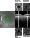
Main body: Parallel mechanisms autoregulate retinal and cerebral microcirculation to maintain blood flow to meet metabolic demands across a range of perfusion pressures. Cerebral blood flow (CBF) is reduced and autoregulation impaired in sepsis, but current methods to image CBF do not reproducibly assess the microcirculation. Peripheral microcirculatory blood flow may be imaged in sublingual and conjunctival mucosa and is impaired in sepsis. Retinal microcirculation can be directly imaged by optical coherence tomography angiography (OCTA) during perfusion-deficit states such as sepsis, and other systemic haemodynamic disturbances such as acute coronary syndrome, and systemic inflammatory conditions such as inflammatory bowel disease.
Conclusion: Monitoring microcirculatory flow offers the potential to enhance monitoring in the care of critically ill patients, and imaging retinal blood flow during critical illness offers a potential biomarker for cerebral microcirculatory perfusion.
Keywords: Critical illness; Optical coherence tomography angiography; Retinal blood flow.
Conflict of interest statement
The authors declare that they have no competing interests.
Figures

References
-
- Trzeciak S, McCoy JV, Phillip Dellinger R, et al. Early increases in microcirculatory perfusion during protocol-directed resuscitation are associated with reduced multi-organ failure at 24 h in patients with sepsis. Intensive Care Med. 2008;34:2210–2217. doi: 10.1007/s00134-008-1193-6. - DOI - PMC - PubMed
Publication types
Grants and funding
LinkOut - more resources
Full Text Sources

