Review
. 2020 Dec;1482(1):61-76.
doi: 10.1111/nyas.14523. Epub 2020 Nov 12.

Advanced endoscopic imaging for detecting and guiding therapy of early neoplasias of the esophagus

Affiliations
Review

Advanced endoscopic imaging for detecting and guiding therapy of early neoplasias of the esophagus

Hiroshi Mashimo et al. Ann N Y Acad Sci. 2020 Dec.

Abstract

Esophageal cancers, largely adenocarcinoma in Western countries and squamous cell cancer in Asia, present a significant burden of disease and remain one of the most lethal of cancers. Key to improving survival is the development and adoption of new imaging modalities to identify early neoplastic lesions, which may be small, multifocal, subsurface, and difficult to detect by standard endoscopy. Such advanced imaging is particularly relevant with the emergence of ablative techniques that often require multiple endoscopic sessions and may be complicated by bleeding, pain, strictures, and recurrences. Assessing the specific location, depth of involvement, and features correlated with neoplastic progression or incomplete treatment may optimize treatments. While not comprehensive of all endoscopic imaging modalities, we review here some of the recent advances in endoscopic luminal imaging, particularly with surface contrast enhancement using virtual chromoendoscopy, highly magnified subsurface imaging with confocal endomicroscopy, optical coherence tomography, elastic scattering spectroscopy, angle-resolved low-coherence interferometry, and light scattering spectroscopy. While there is no single ideal imaging modality, various multimodal instruments are also being investigated. The future of combining computer-aided assessments, molecular markers, and improved imaging technologies to help localize and ablate early neoplastic lesions shed hope for improved disease outcome.

Keywords: Barrett's esophagus; computer-aided diagnosis; confocal laser endomicroscopy; esophageal cancer; narrowband imaging; optical coherence tomography.


Conflict of interest statement

Competing interests:

The authors declare no competing interests.

Figures

Figure 1.
Figure 1.
Examples of NBI in the esophagus. The upper panel shows nondysplastic BE under WLE on the left and corresponding NBI image on the right. The lower panel shows dysplasia in BE under WLE on left and enhanced irregular mucosal pattern under NBI on the right.
Figure 2.
Figure 2.
Endoscopic subsurface imaging tools comparing ultrasonography, OCT/OCM, and confocal microscopy in terms of resolution at the cost of image depth penetration.
Figure 3.
Figure 3.
Examples of CLE images in the human esophagus using intravenous fluorescein contrast. (A) The normal squamous epithelium with intrapapillary capillary loops (IPCL). (B) Specialized intestinal metaplasia (BE). (C) Esophageal adenocarcinoma.
Figure 4.
Figure 4.
Volumetric laser endomicroscopy (VLE) with three views. (A) Looking down from the proximal esophagus. (B) Looking closer to the suspected area of dysplasia. (C) The en face view of the distal esophagus. The luminal en face view shows an area of overlap (yellow arrow) between the three features of dysplasia: orange is a lack of layering, blue is glandular structures, and pink is a hyperreflective surface. Figure and caption were adapted from Trindade et al. and represent the current VLE NinePoint imaging software called Intelligent Real-time Image Segmentation (IRIS)™ artificial intelligence platform.
Figure 5.
Figure 5.
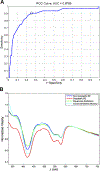
Elastic scattering spectroscopy for detection of the esophageal neoplasm. (A) The receiver−operator curve for sensitivity and specificity in a single-center prospective study. (B) A spectral display of various tissue types in the human esophagus.

Similar articles

Cited by

References

    1. Arnold M, Soerjomataram I, Ferlay J, et al. 2015. Global incidence of oesophageal cancer by histological subtype in 2012. Gut. 64: 381–387. - PubMed
    1. Shaheen NJ, Falk GW, Iyer PG, et al. 2016. ACG Clinical Guideline: Diagnosis and Management of Barrett’s Esophagus. Am J Gastroenterol. 111: 30–50; quiz 51. - PMC - PubMed
    1. Domper Arnal MJ, Ferrandez Arenas A & Lanas Arbeloa A. 2015. Esophageal cancer: Risk factors, screening and endoscopic treatment in Western and Eastern countries. World J Gastroenterol. 21: 7933–7943. - PMC - PubMed
    1. Jemal A, Bray F, Center MM, et al. 2011. Global cancer statistics. CA Cancer J Clin. 61: 69–90. - PubMed
    1. Kandiah K, Chedgy FJ, Subramaniam S, et al. 2017. Early squamous neoplasia of the esophagus: The endoscopic approach to diagnosis and management. Saudi J Gastroenterol. 23: 75–81. - PMC - PubMed

Publication types

MeSH terms

Supplementary concepts