

Fetal brain growth and risk of postnatal white matter injury in critical congenital heart disease
- PMID: 33185192
- PMCID: PMC8012393
- DOI: 10.1016/j.jtcvs.2020.09.096
Fetal brain growth and risk of postnatal white matter injury in critical congenital heart disease
Abstract
Objective: To test the hypothesis that delayed brain development in fetuses with d-transposition of the great arteries or hypoplastic left heart syndrome heightens their postnatal susceptibility to acquired white matter injury.
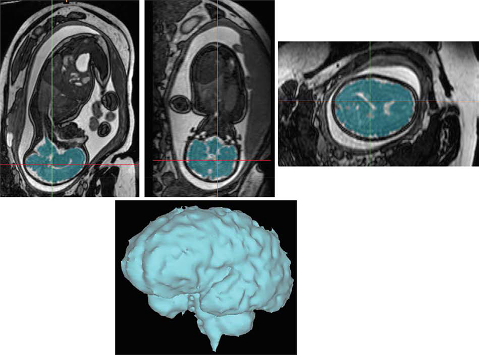
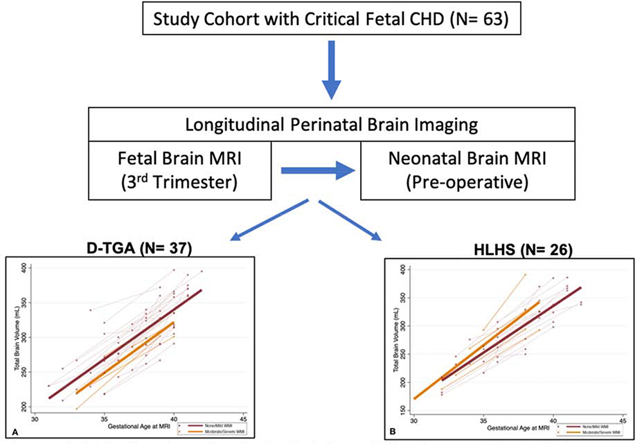
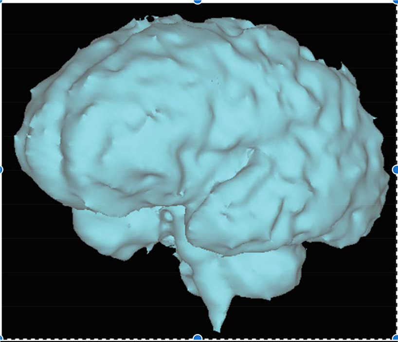
Methods: This is a cohort study across 3 sites. Subjects underwent fetal (third trimester) and neonatal preoperative magnetic resonance imaging of the brain to measure total brain volume as a measure of brain maturity and the presence of acquired white matter injury after birth. White matter injury was categorized as no-mild or moderate-severe based on validated grading criteria. Comparisons were made between the injury groups.
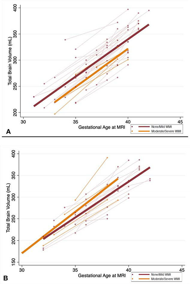
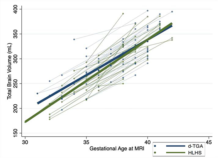
Results: A total of 63 subjects were enrolled (d-transposition of the great arteries: 37; hypoplastic left heart syndrome: 26). White matter injury was present in 32.4% (n = 12) of d-transposition of the great arteries and 34.6% (n = 8) of those with hypoplastic left heart syndrome. Overall total brain volume (taking into account fetal and neonatal scan) was significantly lower in those with postnatal moderate-severe white matter injury compared with no-mild white matter injury after adjusting for age at scan and site in d-transposition of the great arteries (coefficient: 14.8 mL, 95% confidence interval, -28.8 to -0.73, P = .04). The rate of change in total brain volume from fetal to postnatal life did not differ by injury group. In hypoplastic left heart syndrome, no association was noted between overall total brain volume and change in total brain volume with postnatal white matter injury.
Conclusions: Lower total brain volume beginning in late gestation is associated with increased risk of postnatal moderate-severe white matter injury in d-transposition of the great arteries but not hypoplastic left heart syndrome. Rate of brain growth was not a risk factor for white matter injury. The underlying fetal and perinatal physiology has different implications for postnatal risk of white matter injury.
Keywords: brain development; brain injury; congenital heart disease; neurodevelopment.
Copyright © 2020 The American Association for Thoracic Surgery. All rights reserved.
Conflict of interest statement
Figures
Comment in
-
Commentary: The tip of the iceberg: Physiology of congenital cardiac defects and their influence on brain injury.J Thorac Cardiovasc Surg. 2021 Sep;162(3):1020-1021. doi: 10.1016/j.jtcvs.2020.10.004. Epub 2020 Oct 9. J Thorac Cardiovasc Surg. 2021. PMID: 33129502 No abstract available.
-
Commentary: Will fetal brain magnetic resonance imaging guide our timing of surgery for hypoplastic left heart syndrome and transposition of the great arteries?J Thorac Cardiovasc Surg. 2021 Sep;162(3):1018-1019. doi: 10.1016/j.jtcvs.2020.10.109. Epub 2020 Nov 5. J Thorac Cardiovasc Surg. 2021. PMID: 33293068 No abstract available.
-
Commentary: Once again-the heart and the brain.J Thorac Cardiovasc Surg. 2021 Sep;162(3):1017-1018. doi: 10.1016/j.jtcvs.2020.11.079. Epub 2020 Nov 30. J Thorac Cardiovasc Surg. 2021. PMID: 33419541 No abstract available.
-
Commentary: Planning ahead: Fetal magnetic resonance imaging may predict brain injury before surgery for congenital heart disease.J Thorac Cardiovasc Surg. 2021 Sep;162(3):1015-1016. doi: 10.1016/j.jtcvs.2020.11.141. Epub 2020 Dec 14. J Thorac Cardiovasc Surg. 2021. PMID: 33451827 No abstract available.
References
-
- Licht DJ, Wang J, Silvestre DW, Nicolson SC, Montenegro LM, Wernovsky G, et al. Preoperative cerebral blood flow is diminished in neonates with severe congenital heart defects. J Thorac Cardiovasc Surg. 2004December;128(6):841–9. - PubMed
-
- McQuillen PS, Barkovich AJ, Hamrick SEG, Perez M, Ward P, Glidden DV, et al. Temporal and anatomic risk profile of brain injury with neonatal repair of congenital heart defects. Stroke. 2007February;38(2 Suppl):736–41. - PubMed
-
- Miller SP, McQuillen PS, Hamrick S, Xu D, Glidden DV, Charlton N, et al. Abnormal brain development in newborns with congenital heart disease. N Engl J Med. 2007November8;357(19):1928–38. - PubMed
Publication types
MeSH terms
Grants and funding
LinkOut - more resources
Full Text Sources
Medical
