

Development, Screening, and Validation of Camelid-Derived Nanobodies for Neuroscience Research
- PMID: 33185319
- PMCID: PMC7673645
- DOI: 10.1002/cpns.107
Development, Screening, and Validation of Camelid-Derived Nanobodies for Neuroscience Research
Erratum in
-
Corrections.Curr Protoc. 2021 Jul;1(7):e193. doi: 10.1002/cpz1.193. Curr Protoc. 2021. PMID: 34286925 No abstract available.
Abstract
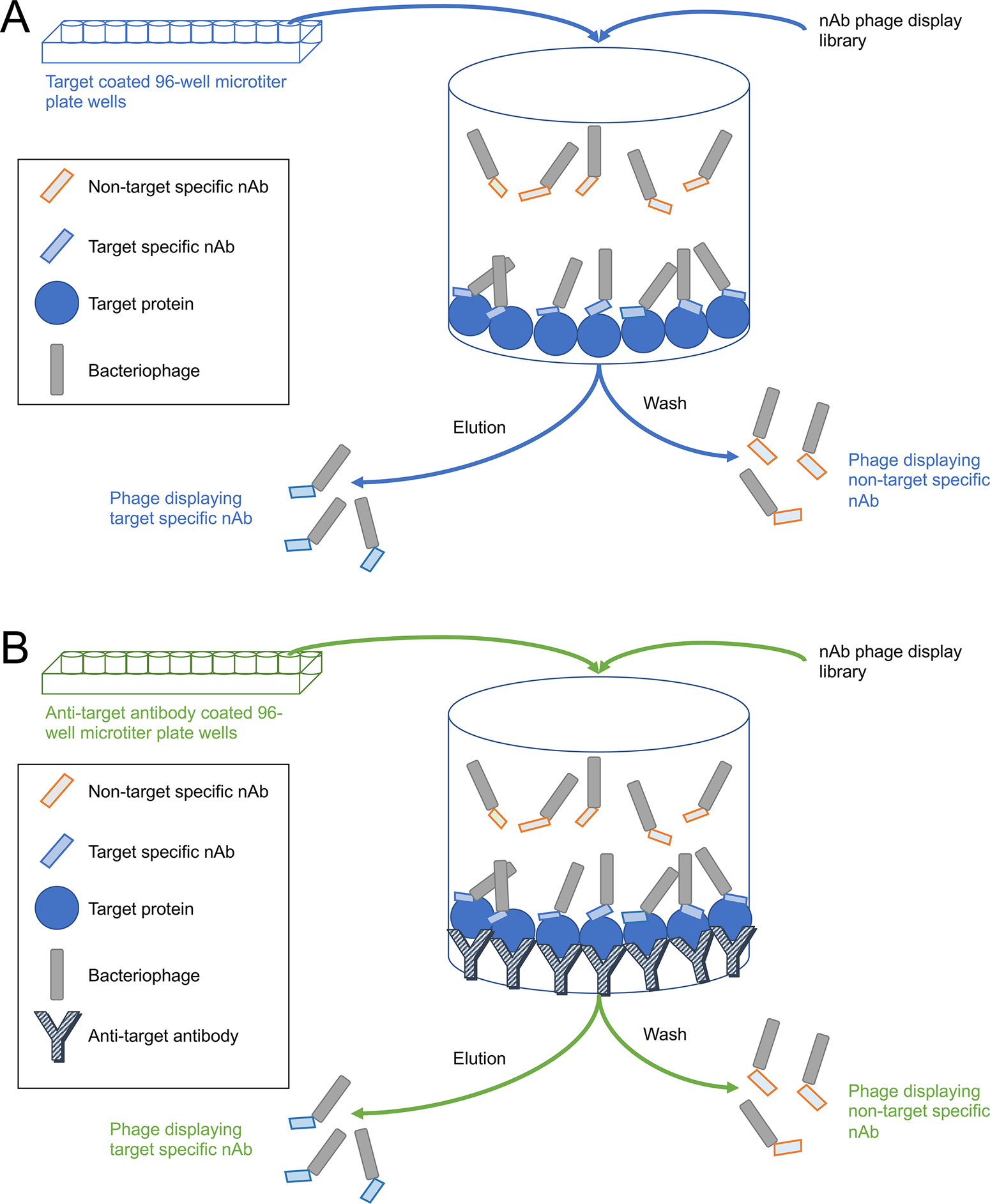
Nanobodies (nAbs) are recombinant antigen-binding variable domain fragments obtained from heavy-chain-only immunoglobulins. Among mammals, these are unique to camelids (camels, llamas, alpacas, etc.). Nanobodies are of great use in biomedical research due to their efficient folding and stability under a variety of conditions, as well as their small size. The latter characteristic is particularly important for nAbs used as immunolabeling reagents, since this can improve penetration of cell and tissue samples compared to conventional antibodies, and also reduce the gap distance between signal and target, thereby improving imaging resolution. In addition, their recombinant nature allows for unambiguous definition and permanent archiving in the form of DNA sequence, enhanced distribution in the form of sequences or plasmids, and easy and inexpensive production using well-established bacterial expression systems, such as the IPTG induction method described here. This article will review the basic workflow and process for developing, screening, and validating novel nAbs against neuronal target proteins. The protocols described make use of the most common nAb development method, wherein an immune repertoire from an immunized llama is screened via phage display technology. Selected nAbs can then be taken through validation assays for use as immunolabels or as intrabodies in neurons. © 2020 Wiley Periodicals LLC. Basic Protocol 1: Total RNA isolation from camelid leukocytes Basic Protocol 2: First-strand cDNA synthesis; VH H and VH repertoire PCR Basic Protocol 3: Preparation of the phage display library Basic Protocol 4: Panning of the phage display library Basic Protocol 5: Small-scale nAb expression Basic Protocol 6: Sequence analysis of selected nAb clones Basic Protocol 7: Nanobody validation as immunolabels Basic Protocol 8: Generation of nAb-pEGFP mammalian expression constructs Basic Protocol 9: Nanobody validation as intrabodies Support Protocol 1: ELISA for llama serum testing, phage titer, and screening of selected clones Support Protocol 2: Amplification of helper phage stock Support Protocol 3: nAb expression in amber suppressor E. coli bacterial strains.
Keywords: ELISA; immunocytochemistry; immunohistochemistry; intrabodies; nanobodies; phage display; recombinant antibodies.
© 2020 Wiley Periodicals LLC.
Conflict of interest statement
Conflict of Interest
The authors have no conflict of interest to declare.
Figures

References
-
- Andris-Widhopf J, Rader C, Steinberger P, Fuller R, & Barbas CF 3rd. (2000). Methods for the generation of chicken monoclonal antibody fragments by phage display. J Immunol Methods, 242(1–2), 159–181. - PubMed
Publication types
MeSH terms
Substances
Grants and funding
LinkOut - more resources
Full Text Sources
Miscellaneous
