. 2020 Nov 13:9:e58277.
doi: 10.7554/eLife.58277.

Bacterial OTU deubiquitinases regulate substrate ubiquitination upon Legionella infection

Affiliations

Bacterial OTU deubiquitinases regulate substrate ubiquitination upon Legionella infection

Donghyuk Shin et al. Elife. .

Abstract

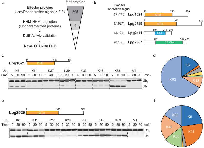
Legionella pneumophila causes a severe pneumonia known as Legionnaires' disease. During the infection, Legionella injects more than 300 effector proteins into host cells. Among them are enzymes involved in altering the host-ubiquitination system. Here, we identified two LegionellaOTU (ovarian tumor)-like deubiquitinases (LOT-DUBs; LotB [Lpg1621/Ceg23] and LotC [Lpg2529]). The crystal structure of the LotC catalytic core (LotC14-310) was determined at 2.4 Å. Unlike the classical OTU-family, the LOT-family shows an extended helical lobe between the Cys-loop and the variable loop, which defines them as a unique class of OTU-DUBs. LotB has an additional ubiquitin-binding site (S1'), which enables the specific cleavage of Lys63-linked polyubiquitin chains. By contrast, LotC only contains the S1 site and cleaves different species of ubiquitin chains. MS analysis of LotB and LotC identified different categories of host-interacting proteins and substrates. Together, our results provide new structural insights into bacterial OTU-DUBs and indicate distinct roles in host-pathogen interactions.

Keywords: Legionella pneumophila; OTU-deubiquitinase; bacterial deubiquitinase; biochemistry; chemical biology; effector proteins; human; ubiquitin.


Conflict of interest statement

DS, AB, YC, MA, AM, Gv, HO, GH No competing interests declared, ID Reviewing editor, eLife

Figures

Figure 1.
Figure 1.. Identification of novel deubiquitinases (DUBs) in Legionella pneumophila.
(a) Graphical illustration of identification of novel DUBs from L. pneumophila effector proteins. (b) Predicted DUB domain of four putative Legionella DUBs. (c, e) Time-course di-ubiquitin panel cleavage assay with Lpg1621 (LotB) and Lpg2529 (LotC). (d, f) Linkage specificity diagram of Lpg1621 (LotB) and Lpg2529 (LotC). The percentage of cleaved ubiquitin species at 90 min was plotted.
Figure 1—figure supplement 1.
Figure 1—figure supplement 1.. Ubiquitin cleavage assay with putative deubiquitinases (DUBs) from Legionella.
(a) Di-ubiquitin species were incubated with putative Legionella DUBs and analyzed by immuno-blotting with ubiquitin antibody. (b) HEK293 cell lysates were treated with purified Legionella DUBs and analyzed by immuno-blotting with indicated antibodies.
Figure 2.
Figure 2.. Biochemical properties of LotB and LotC.
(a) Predicted catalytic residues on LotB and LotC. (b, c) Di-Ub cleavage activity assay with wild-type and catalytic mutants of LotB and LotC. (d, e) Activity-based probes (ABPs) test on LotB and LotC. Propargyl-Ub-ABP (Prg-ABP) and vinylmethylester-ubiquitin-ABP (VME-ABP) were incubated as indicated time-points with LotB and LotC and analyzed on SDS-PAGE with coomassie staining. (f, g) Propargyl ubiquitin or ubiquitin-like modifiers reactivity test on LotB and LotC. Prg-ABPs are incubated with LotB and LotC with indicated time points.
Figure 2—figure supplement 1.
Figure 2—figure supplement 1.. Biochemical properties of LotB and LotC.
(a, b) Propargyl (Prg) – ubiquitin or ubiquitin-like modifiers reactivity test on LotB and LotC. Prg-ABPs are incubated with LotB and LotC for 60 min. (c) Sequence alignment of ubiquitin with NEDD8. Non-conserved residues are marked with red-reversed triangle.
Figure 3.
Figure 3.. Structural comparison of Legionella OTU-deubiquitinases with other OTU-family.
(a, b) Minimal domain boundaries of catalytically active LotB and LotC. Different constructs were cloned based on the predicted OTU-domains and their activity, and were tested with di-Ub panel. (c, d) Structural comparison of LotB and LotC with the closest homologues. CCHF- (PDB: 3PHU), OTUD2 (PDB: 4BOQ), OTUD3 (PDB: 4BOU), Taggert- (PDB: 6D × 3), DGK nairo- (PDB: 6D × 2), Otubain1 (PDB: 2ZFY), Otubain2 (PDB: 4FJV). (e) Sequence alignment of LotB and LotC with their closest homologues. Catalytic cysteine and histidine are highlighted in red and conserved residues are highlighted in yellow.
Figure 3—figure supplement 1.
Figure 3—figure supplement 1.. Sequence alignment of OTU deubiquitinase family.
The helical lobe between the catalytic Cys loop and the variable loop is shown as bar. The catalytic Cys is highlighted in red and conserved residues are highlighted in yellow.
Figure 4.
Figure 4.. Ubiquitin-binding sites on LotB and LotC.
(a, b) Molecular docking and simulations of monoubiquitin to LotB and LotC. Shown are representative snapshots of the MD simulations. Catalytic cysteine and key residues for the interaction between ubiquitin and LotB or LotC are depicted as sticks. (c, d) Key residues mediating interactions between ubiquitin and LotB or LotC. Residues are highlighted in the structure (left). Side-chain center-of-mass distances are shown as a function of the simulation time (right). (e, f) Di-ubiquitin cleavage assay for mutants of LotB and LotC. The catalytic activity of LotB or LotC wild-type and their mutants was tested with K63- or K48-linked Ub2, respectively.
Figure 5.
Figure 5.. Host-interacting proteins and cellular localization of LotB and LotC.
(a, c) Proteomic analysis of interacting partners of LotB and LotC. Catalytically inactive FLAG-LotB (C29A) and FLAG-LotC (C24A) were transfected and immunoprecipitated. Co-precipitated interacting proteins were analyzed by mass spectrometry. (b, d) Cellular localization of LotB and LotC. FLAG-tagged LotB and LotC were ectopically expressed in U2OS cells and immune-stained with cellular organelle markers (endoplasmic reticulum: Calnexin, mitochondria: TOMM20, Golgi: GM130).
Figure 5—figure supplement 1.
Figure 5—figure supplement 1.. Cellular localization of LotB full-length and LotB-OTU.
FLAG-tagged LotB or LotC-OTU catalytic core was ectopically expressed in U2OS cells and immune-stained with cellular organelle markers (endoplasmic reticulum: Calnexin, mitochondria: TOMM20, Golgi: GM130).
Figure 5—figure supplement 2.
Figure 5—figure supplement 2.. Proteomic analysis of interacting partners of LotB and LotC together with Legionella E3s.
(a, b) Proteomic analysis of interacting partners of LotB together with SdcA and SidC, respectively. Catalytically inactive FLAG-LotB (C29A) was co-transfected with either SdcA or SidC and immunoprecipitated. Co-precipitated interacting proteins were analyzed by mass spectrometry. (c, d) Proteomic analysis of interacting partners of LotC together with SdcA and SidC, respectively. Catalytically inactive FLAG-LotC (C24A) was co-transfected with either SdcA or SidC and immunoprecipitated. Co-precipitated interacting proteins were analyzed by mass spectrometry.
Figure 6.
Figure 6.. Substrate identification of LotB and LotC proteomic analysis of potential substrates of LotB and LotC.
(a–c) Schematic of the experiment and subsequent validation using western blot. (d, e) Volcano plot depicting the identified proteins with corresponding fold change and p-values. Comparison was done between Mut and WT deubiquitinase (DUB). Enriched proteins with Log2 Fold change ≥ 0.5 along with −Log10 p-value ≥ 1.3 was considered for further validation. (f–h) Immunoprecipitation of myc from the infected lysates was performed to enrich the potential substrates for LotB, which are RYK, Rab13, and PCYT1A, and for LotC, which are VAT1, HMOX1, and PPP2R1A, respectively. The enriched potential substrates were further incubated with wild-type or catalytic dead mutant DUB, followed by western blotting to detect ubiquitin and myc expression.

Similar articles

Cited by

References

    1. Abdul Rehman SA, Kristariyanto YA, Choi SY, Nkosi PJ, Weidlich S, Labib K, Hofmann K, Kulathu Y. MINDY-1 is a member of an evolutionarily conserved and structurally distinct new family of deubiquitinating enzymes. Molecular Cell. 2016;63:146–155. doi: 10.1016/j.molcel.2016.05.009. - DOI - PMC - PubMed
    1. Abraham MJ, Murtola T, Schulz R, Páll S, Smith JC, Hess B, Lindahl E. GROMACS: high performance molecular simulations through multi-level parallelism from laptops to supercomputers. SoftwareX. 2015;1-2:19–25. doi: 10.1016/j.softx.2015.06.001. - DOI
    1. Afonine PV, Grosse-Kunstleve RW, Echols N, Headd JJ, Moriarty NW, Mustyakimov M, Terwilliger TC, Urzhumtsev A, Zwart PH, Adams PD. Towards automated crystallographic structure refinement with phenix.refine. Acta Crystallographica Section D Biological Crystallography. 2012;68:352–367. doi: 10.1107/S0907444912001308. - DOI - PMC - PubMed
    1. Akutsu M, Ye Y, Virdee S, Chin JW, Komander D. Molecular basis for ubiquitin and ISG15 cross-reactivity in viral ovarian tumor domains. PNAS. 2011;108:2228–2233. doi: 10.1073/pnas.1015287108. - DOI - PMC - PubMed
    1. Best RB, Zhu X, Shim J, Lopes PE, Mittal J, Feig M, Mackerell AD. Optimization of the additive CHARMM all-atom protein force field targeting improved sampling of the backbone φ, ψ and side-chain χ(1) and χ(2) dihedral angles. Journal of Chemical Theory and Computation. 2012;8:3257–3273. doi: 10.1021/ct300400x. - DOI - PMC - PubMed

Publication types

Substances

LinkOut - more resources