Review
. 2022 Feb 24;65(4):2716-2746.
doi: 10.1021/acs.jmedchem.0c01140. Epub 2020 Nov 13.

Targeting SARS-CoV-2 Proteases and Polymerase for COVID-19 Treatment: State of the Art and Future Opportunities

Affiliations
Review

Targeting SARS-CoV-2 Proteases and Polymerase for COVID-19 Treatment: State of the Art and Future Opportunities

Rolando Cannalire et al. J Med Chem. .

Abstract

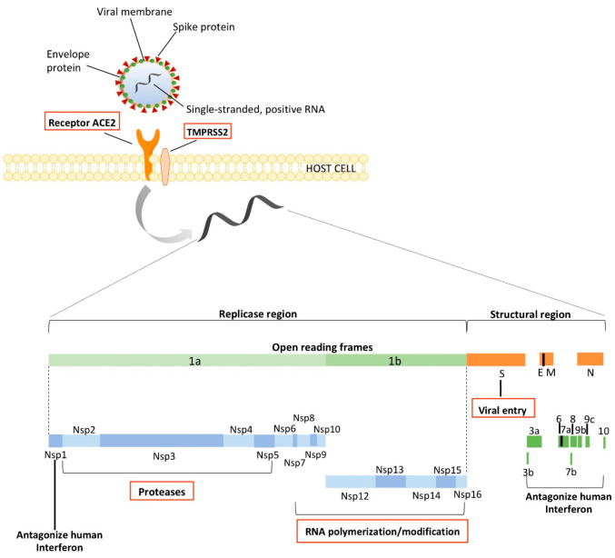
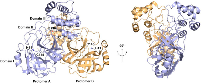
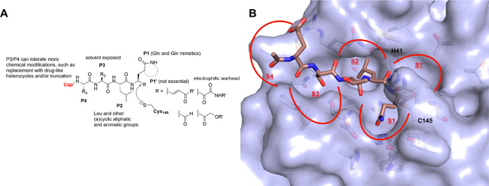
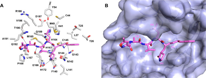
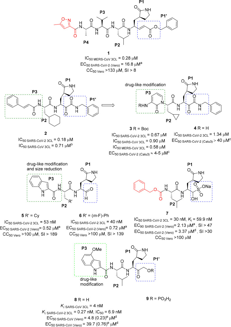
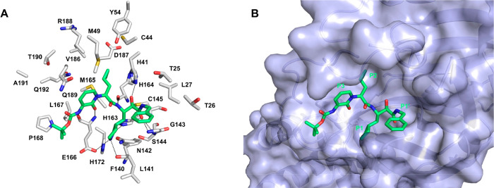
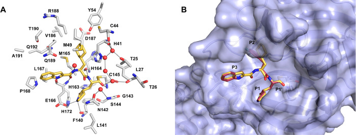
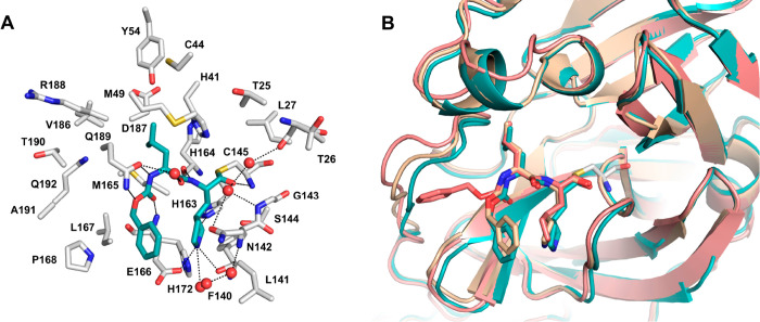
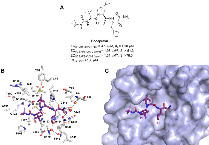
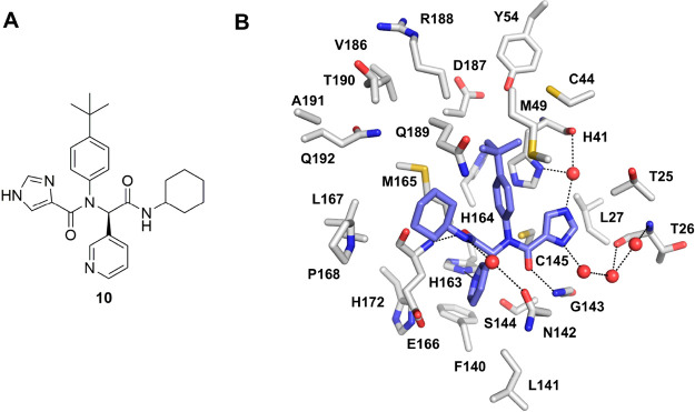
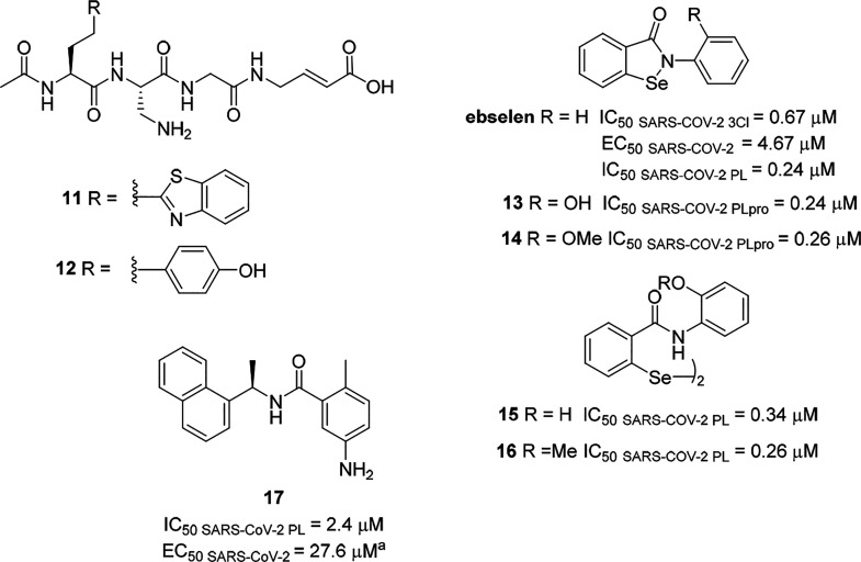
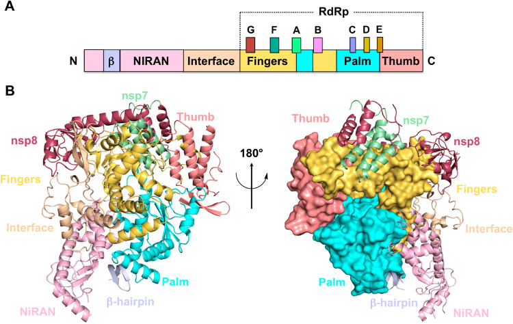
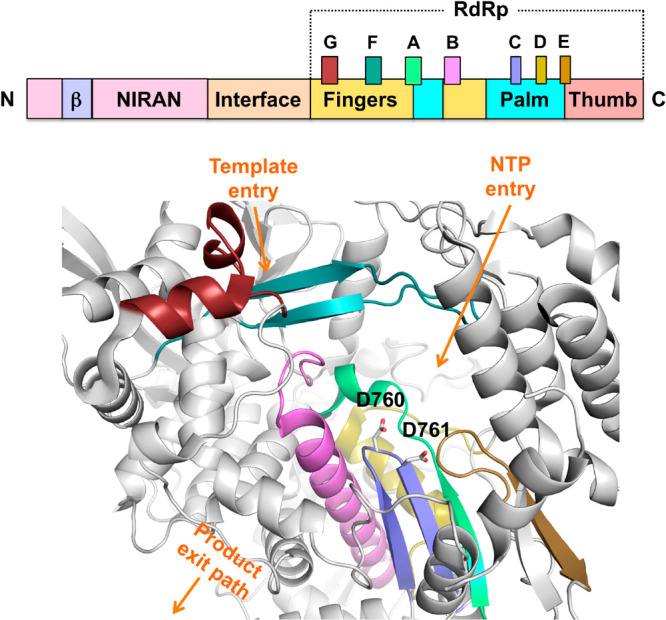
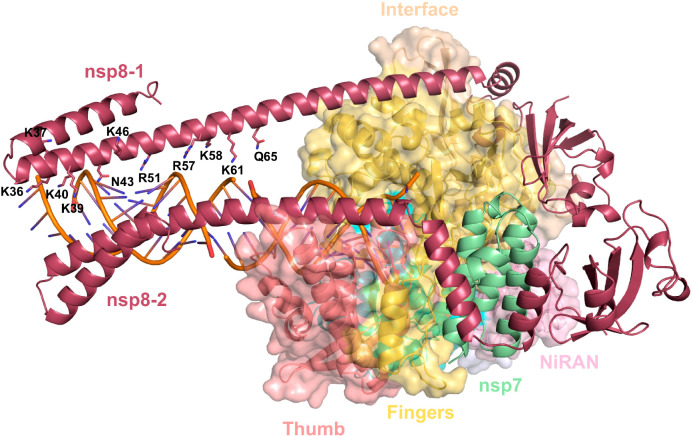
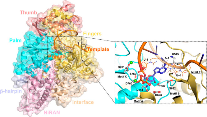
The newly emerged coronavirus, called SARS-CoV-2, is the causing pathogen of pandemic COVID-19. The identification of drugs to treat COVID-19 and other coronavirus diseases is an urgent global need, thus different strategies targeting either virus or host cell are still under investigation. Direct-acting agents, targeting protease and polymerase functionalities, represent a milestone in antiviral therapy. The 3C-like (or Main) protease (3CLpro) and the nsp12 RNA-dependent RNA-polymerase (RdRp) are the best characterized SARS-CoV-2 targets and show the highest degree of conservation across coronaviruses fostering the identification of broad-spectrum inhibitors. Coronaviruses also possess a papain-like protease, another essential enzyme, still poorly characterized and not equally conserved, limiting the identification of broad-spectrum agents. Herein, we provide an exhaustive comparative analysis of SARS-CoV-2 proteases and RdRp with respect to other coronavirus homologues. Moreover, we highlight the most promising inhibitors of these proteins reported so far, including the possible strategies for their further development.


Conflict of interest statement

The authors declare no competing financial interest.

Figures

Figure 1
Figure 1
Schematic representation of SARS-CoV-2 virion, viral entry, genome translation, and polyprotein processing.
Figure 2
Figure 2
Overview of SARS-CoV-2 3CLpro architecture. The X-ray structure of SARS-CoV-2 3CLpro (PDB 6Y2G) is shown as a ribbon model in two different orientations. For clarity, the bound inhibitor has been removed. Protomers A (light-blue) and B (light-orange) associate into a dimer stabilized by a salt bridge between Glu290 and Arg4, while the substrate binding site resides at the interface of domains I and II. The catalytic residues Cys145 and His41 are highlighted.
Figure 3
Figure 3
Schematization of the main features of 3CLpro inhibitors and protease subsite specificity. (A) General representation of peptidic covalent reversible inhibitors of SARS-CoV-2 3CLpro summarizing the chemical requirements and SAR of compounds so far reported in literature. (B) Surface representation of the active site pocket of SARS-CoV 3CLpro bound to a peptide aldehyde inhibitor (dark salmon sticks, PDB 3SNE), chosen as a representative substrate-like inhibitor. The S1–S4 and S1′ subsites are indicated with red lines and labeled. The key residues forming the active site pocket are displayed as white sticks; the catalytic residues Cys145 and His41 are labeled.
Figure 4
Figure 4
Co-crystallographic pose of compound 1 (violet sticks, PDB 7BQY) covalently bound to the active site of SARS-CoV-2 3CLpro. (A) The key residues forming the active site pocket are displayed as white sticks and labeled. H-bonds are depicted as dashed black lines. (B) Surface representation of the active site pocket with bound N3. The P1–P4 and P1′ moieties are labeled.
Figure 5
Figure 5
Compounds 19 with biological activities. aAntiviral activity evaluated by viral plaque assay. bData from ref (42). cAntiviral activity evaluated by viral RNA qRT-PCR quantification. dAntiviral activity evaluated by CPE reduction. eValues in bracket have been obtained in the presence of a P-gp inhibitor. The main structural differences, significant modifications, and warheads are highlighted.
Figure 6
Figure 6
Co-crystallographic pose of compound 3 (green sticks, PDB 6Y2F) covalently bound to the active site of SARS-CoV-2 3CLpro. (A) The key residues forming the active site pocket are displayed as white sticks. H-bonds are depicted as dashed black lines. (B) Surface representation of the active site pocket with bound compound 3.
Figure 7
Figure 7
Co-crystallographic pose of compound 5 (yellow-orange sticks, PDB 6LZE) covalently bound to the active site of SARS-CoV-2 3CLpro. (A) The key residues forming the binding pocket are displayed as white sticks; water molecules are shown as red spheres. H-bonds are depicted as dashed black lines. (B) Overlay of 5 and 6 (raspberry sticks, PDB 6M0K) co-crystallographic poses.
Figure 8
Figure 8
Co-crystallographic pose of compound 7 (teal sticks, PDB 7BRR) covalently bound to the active site of SARS-CoV-2 3CLpro. (A) The key residues forming the active site pocket are displayed as white sticks; water molecules are shown as red spheres. H-bonds are depicted as dashed black lines. (B) Overlay of 7 bound to SARS-CoV-2 3CLpro structures (PDBs: 7BRR, teal; 6WTJ, wheat; 6WTT, salmon), showing the different conformations of the benzyloxycarbonyl group.
Figure 9
Figure 9
(A) Structure and biological activity of Boceprevir. aAntiviral activity evaluated by viral plaque assay. bAntiviral activity evaluated by CPE reduction. (B) Co-crystallographic pose of Boceprevir (purple sticks, PDB 6WNP) covalently bound to the active site of SARS-CoV-2 3CLpro. The key residues forming the active site pocket are displayed as white sticks; water molecules are displayed as red spheres. H-bonds are depicted as dashed black lines. (C) Surface representation of the active site pocket with bound Boceprevir.
Figure 10
Figure 10
(A) Structures and biological activities of flavones Baicalein and Baicalin. aAntiviral activity evaluated by viral RNA measurement by qRT-PCR. (B) Co-crystallographic pose of Baicalein (light-pink sticks, PDB 6M2N) into the active site of SARS-CoV-2 3CLpro. The key residues forming the binding pocket are displayed as white sticks. The buried water molecule is displayed as a red sphere. H-bonds are depicted as dashed black lines.
Figure 11
Figure 11
(A) Chemical structure of compound 10. (B) Co-crystallographic pose of 10 (slate sticks, PDB 6W63) into the active site of SARS-CoV-2 3CLpro. The key residues forming the binding pocket are displayed as white sticks; water molecules are displayed as red spheres. H-bonds are depicted as dashed black lines.
Figure 12
Figure 12
Chemical structures and biological activities of Ebselen and compounds 1117.
Figure 13
Figure 13
Architecture of SARS-CoV-2 nsp12. (A) Schematic diagram outlining the domain organization of SARS-CoV-2 nsp12. The domains are colored as: palm, cyan; fingers, yellow-orange; thumb, salmon. The N-terminal NiRAN domain, the interface and the β-hairpin are colored light-pink, wheat, and blue-white, respectively. The polymerase conserved motifs are colored as: motif A, lime green; motif B, violet; motif C, slate; motif D, olive; motif E, sand; motif F, deep-teal; motif G, ruby. (B) Structure of SARS-CoV-2 in two different orientations (PDB 6M71). (left) Ribbon diagram of nsp12 showing the arrangement of palm, fingers, and thumb domains. Domains are colored as in (A). Nsp7 and nsp8 cofactors are shown as pale-green and raspberry ribbon models, respectively. (right) The palm, fingers, and thumb domains are shown as a molecular surface.
Figure 14
Figure 14
Overview of SARS-CoV-2 nsp12 active site. For clarity, the RdRp core region is shown as a white ribbon model, whereas the polymerase conserved motifs (A–G) are colored according to the upper schematic diagram. The catalytic residues Asp760 and Asp761 are shown as white sticks. The template entry, NTP entry, product hybrid exits paths are indicated by orange arrows.
Figure 15
Figure 15
Structure of SARS-CoV-2 replicating RdRp-RNA complex (PDB 6YYT). Nsp12 is shown as a molecular surface (color code as in Figure 13), whereas the cofactors nsp7 and nsp8 (protomers 1 and 2) are shown as pale-green and raspberry ribbon models, respectively. RNA turns are shown as an orange ribbon model. The positively charged nsp8 residues, proposed to interact with RNA, are shown as sticks.
Figure 16
Figure 16
Putative mechanism of ProTides in vivo metabolism. Upon diffusion into the cell, the amino acid ester of the ProTide is cleaved by intracellular esterases, then a cyclization occurs onto the phosphorus, with the release of the phenoxide moiety. The unstable cyclic intermediate is then hydrolyzed by water to the alanine metabolite, whose P–N bond is hydrolyzed by phosphoramidase-type enzymes to unmask the NI monophosphate form. The NI monophosphate is routed to further phosphorylation steps, yielding the active triphosphate form (NTP) and thus circumventing the endogenous phosphorylation pathway (orange box). X, aromatic substituents; Y, O, or CH2; R, ester substituents.
Figure 17
Figure 17
Chemical structures of antiviral nucleoside analogues in clinical development. The parent bases are highlighted in light-blue (adenine), green (guanine), and orange (cytosine).
Figure 18
Figure 18
Schematization of Remdesivir metabolic conversion to the active triphosphate form. Remdesivir is first transformed into the intermediate alanine metabolite 20, then to the monophosphate form 21 and finally to the triphosphate form 22. The phosphoramidate prodrug moiety is shown in magenta. 19 (GS-441524) is the parent nucleoside of Remdesivir.
Figure 19
Figure 19
Binding mode of Remdesivir into the SARS-CoV-2 nsp12 active site (PDB 7BV2). (left) nsp12 is shown as a molecular surface, colored according to the schematic diagram in Figure 13. For clarity, nsp7 and nsp8 cofactor have been removed. The template and primer RNA are shown as ribbon models and labeled. (right) Zoom-in of the nsp12 active site. The covalently bound monophosphate form of Remdesivir (slate) and the pyrophosphate group are shown as sticks. Magnesium ions are shown as green spheres. The RNA bases interacting with Remdesivir are shown as orange thin sticks, while protein residues are shown as white thick sticks. Hydrogen bonds are shown as black dashed lines.
Figure 20
Figure 20
A close view of the covalently bound Remdesivir within the RdRp active site. (A) Remdesivir is incorporated into the primer strand and terminates chain elongation (PDB 7BV2). (B) Remdesivir incorporation induces a mechanism of delayed-chain termination (PDB 7C2K). RNA is shown as orange ribbon model. For clarity, only key residues (thick white sticks) and bases (orange thin sticks) that interact with Remdesivir are shown. Hydrogen bonds are shown as dashed lines.
Figure 21
Figure 21
Schematization of Favipiravir metabolic conversion. Favipiravir is first converted to its ribofuranosyl monophosphate derivative 23 and subsequently to the triphosphate active form 24.
Figure 22
Figure 22
Chemical structures of the prodrug 18 and its parent compound 25. The isobutyrate ester prodrug moiety is shown in blue.

Similar articles

Cited by

References

    1. Ten Threats to Global Health in 2019; World Health Organization, 2019; https://www.who.int/emergencies/ten-threats-to-global-health-in-2019 (accessed 2020-09-28).
    1. Prioritizing Diseases for Research and Development in Emergency Contexts; World Health Organization, 2020; https://www.who.int/activities/prioritizing-diseases-for-research-and-de... (accessed 2020-09-28).
    1. Wu F.; Zhao S.; Yu B.; Chen Y. M.; Wang W.; Song Z. G.; Hu Y.; Tao Z. W.; Tian J. H.; Pei Y. Y.; Yuan M. L.; Zhang Y. L.; Dai F. H.; Liu Y.; Wang Q. M.; Zheng J. J.; Xu L.; Holmes E. C.; Zhang Y. Z. A New Coronavirus Associated with Human Respiratory Disease in China. Nature 2020, 579, 265–269. 10.1038/s41586-020-2008-3. - DOI - PMC - PubMed
    1. Zhu N.; Zhang D.; Wang W.; Li X.; Yang B.; Song J.; Zhao X.; Huang B.; Shi W.; Lu R.; Niu P.; Zhan F.; Ma X.; Wang D.; Xu W.; Wu G.; Gao G. F.; Tan W. A Novel Coronavirus from Patients with Pneumonia in China, 2019. N. Engl. J. Med. 2020, 382, 727–733. 10.1056/NEJMoa2001017. - DOI - PMC - PubMed
    1. Coronavirus (COVID-19) Events As They Happen; World Health Organisation, 2019; https://www.who.int/emergencies/diseases/novel-coronavirus-2019/events-a... (accessed 2020-09-28).

MeSH terms