. 2020 Nov 13;15(11):e0240197.
doi: 10.1371/journal.pone.0240197. eCollection 2020.

Tuning cell behavior with nanoparticle shape

Affiliations

Tuning cell behavior with nanoparticle shape

Edoardo Scarpa et al. PLoS One. .

Abstract

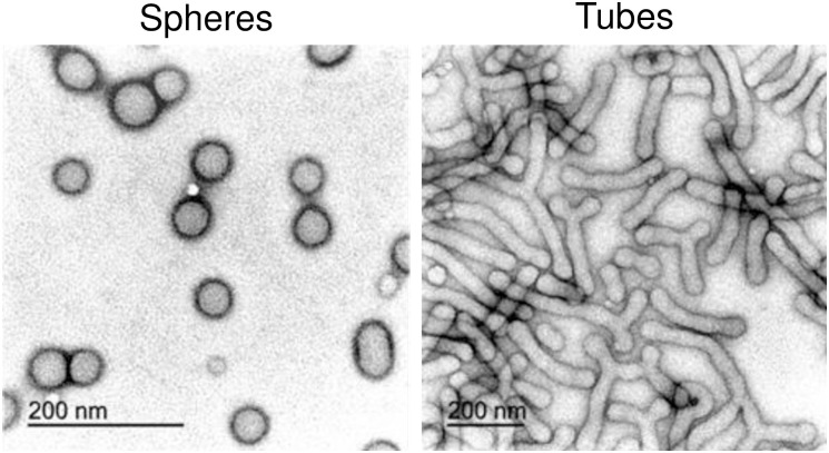
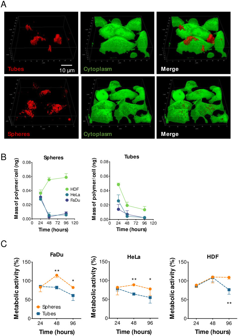
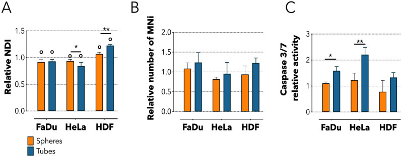
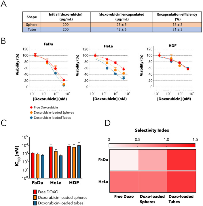
We investigated how the shape of polymeric vesicles, made by the exact same material, impacts the replication activity and metabolic state of both cancer and non-cancer cell types. First, we isolated discrete geometrical structures (spheres and tubes) from a heterogeneous sample using density-gradient centrifugation. Then, we characterized the cellular internalization and the kinetics of uptake of both types of polymersomes in different cell types (either cancer or non-cancer cells). We also investigated the cellular metabolic response as a function of the shape of the structures internalized and discovered that tubular vesicles induce a significant decrease in the replication activity of cancer cells compared to spherical vesicles. We related this effect to the significant up-regulation of the tumor suppressor genes p21 and p53 with a concomitant activation of caspase 3/7. Finally, we demonstrated that combining the intrinsic shape-dependent effects of tubes with the delivery of doxorubicin significantly increases the cytotoxicity of the system. Our results illustrate how the geometrical conformation of nanoparticles could impact cell behavior and how this could be tuned to create novel drug delivery systems tailored to specific biomedical application.


Conflict of interest statement

We acknowledge Astrazeneca for covering part of the S.C.D.S. salary. We also thank British Technology Group (BTG) for donating the MPC monomer. There are no patents, products in development or marketed products to declare. This does not alter our adherence to PLOS ONE policies on sharing data and materials.

Figures

Fig 1
Fig 1. TEM characterization of spherical and tubular polymersomes.
Transmission electron microscopy micrographs showing the morphologies of the polymersomes following preparation and purification using sucrose-based density gradient centrifugation.
Fig 2
Fig 2. Cellular uptake of spheres and tubes.
(A) Confocal imaging showing the complete internalization of spheres after 96 hours incubation with FaDu cells and the “frustrated” uptake of the tubes. Green is Calcein staining of the cytoplasm. (B) HPLC-based quantification of the mass of polymer per cell over time in either FaDu, HeLa or HDF cells. (C) Viability assay on FaDu, HeLa, or HDF cells incubated with spheres or tubes for 24, 48 or 96 hours. Two-way ANOVA. * = p < 0.05, ** = p < 0.01. n = 3 independent experiments.
Fig 3
Fig 3. Cellular metabolic effects following spheres or tubes internalization.
(A) Nuclear Division Index (NDI) quantification for FaDu, HeLa and HDF cells after 24 hours incubation with spheres or tubes. (B) Micronucleus assay (MNi) for addressing the presence of DNA damage after 24 hours incubation with spheres or tubes. (C) Caspase 3/7 assay for analyzing the activation of extrinsic apoptosis following 24 hours incubation with spheres or tubes. Two-way ANOVA. * = p < 0.05, ** = p < 0.01, o = statically different compared to the untreated control. n = 3 independent experiments.
Fig 4
Fig 4. Cellular metabolic effects following spheres or tubes internalization.
(A) Real Time qPCR for quantifying the expression of genes involved in replication activity (p21, p53) following 24 hours incubation with spheres or tubes. Two-way ANOVA. * = p < 0.05, ** = p < 0.01, *** = p < 0.001, **** = p < 0.0001; o = statically different compared to the untreated control. n = 3 independent experiments. (B) Heat map showing real time qPCR quantification of genes involved in oxidative stress (SOD1, CAT), detoxification metabolism (CYP1A1, CYP1B1), Unfolded Protein Response—UPR (ATF4, ATF6), and general shock (HSP27, HSP70). Cells were incubated for following 24 hours incubation with spheres or tubes. n = 3 independent experiments.
Fig 5
Fig 5. Enhanced cytotoxic activity of doxorubicin-loaded tubes.
(A) Table summarizing the initial mass of doxorubicin solubilized with the block co-polymer, the final concentration encapsulated as measured by HPLC and the overall efficiency of encapsulation. (B) Cell viability test on HDF, HeLa and FaDu cells following 48 hours incubation with increasing extracellular concentrations of either free doxorubicin or doxorubicin loaded in spheres or tubes. (C) IC50 values (nM) for the free (green), spheres-encapsulated (orange) and tubes-encapsulated (blue) DOXO. (D) Heat-map showing the selectivity index (SI) of free doxorubicin, doxorubicin-loaded spheres, or doxorubicin-loaded tubes towards FaDu or HeLa cells. Note that SI is the log ratio between the IC50 in non-cancer and in cancer cells (HDF as reference).

References

    1. Toy R, Peiris PM, Ghaghada KB, Karathanasis E. Shaping cancer nanomedicine: The effect of particle shape on the in vivo journey of nanoparticles Nanomedicine. Future Medicine Ltd.; 2014. pp. 121–134. 10.2217/nnm.13.191 - DOI - PMC - PubMed
    1. Park KH, Chhowalla M, Iqbal Z, Sesti F. Single-walled carbon nanotubes are a new class of ion channel blockers. J Biol Chem. 2003. 10.1074/jbc.M310216200 - DOI - PubMed
    1. Andón FT, Fadeel B. Programmed Cell Death: Molecular Mechanisms and Implications for Safety Assessment of Nanomaterials. Acc Chem Res. 2012. 10.1021/ar300020b - DOI - PubMed
    1. Leifert A, Pan Y, Kinkeldey A, Schiefer F, Setzler J, Scheel O, et al. Differential hERG ion channel activity of ultrasmall gold nanoparticles. Proc Natl Acad Sci. 2013. 10.1073/pnas.1220143110 - DOI - PMC - PubMed
    1. Ruiz-Perez L, Messager L, Gaitzsch J, Joseph A, Sutto L, Gervasio FL, et al. Molecular engineering of polymersome surface topology. Sci Adv. 2016;2: e1500948–e1500948. 10.1126/sciadv.1500948 - DOI - PMC - PubMed

Publication types