. 2020 Nov 2;61(13):19.
doi: 10.1167/iovs.61.13.19.

Measuring the Contributions of Basal Laminar Deposit and Bruch's Membrane in Age-Related Macular Degeneration

Affiliations

Measuring the Contributions of Basal Laminar Deposit and Bruch's Membrane in Age-Related Macular Degeneration

Amol A Sura et al. Invest Ophthalmol Vis Sci. .

Abstract

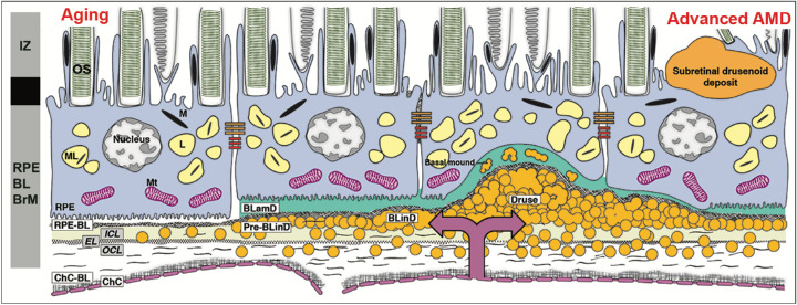
Purpose: Basal laminar deposit (BLamD) is a consistent finding in age-related macular degeneration (AMD). We quantified BLamD thickness, appearance, and topography in eyes of aged donors with and without AMD and evaluated its relationship to other components of the retinal pigment epithelium-basal lamina/Bruch's membrane (RPE-BL-BrM) complex.

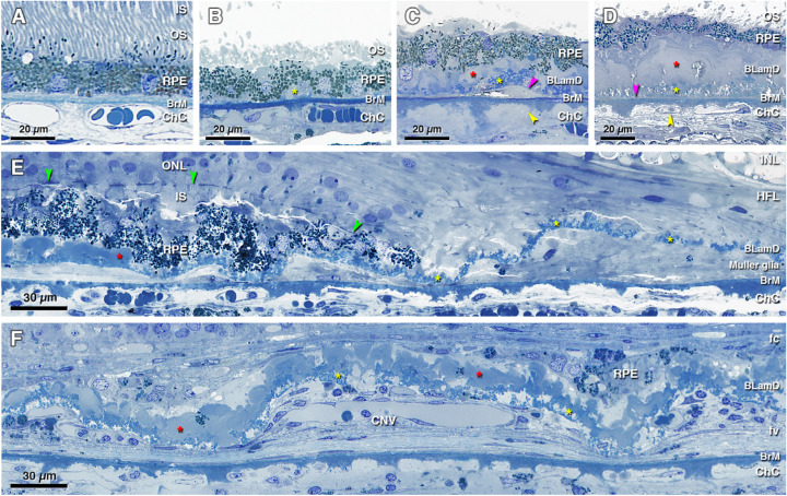
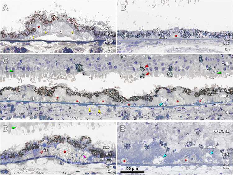
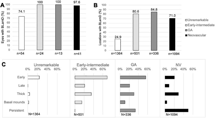
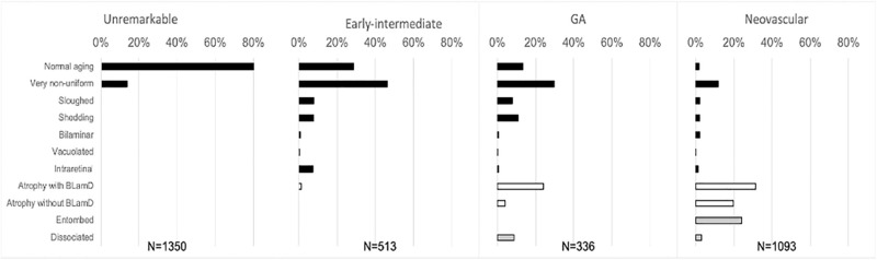
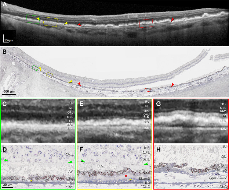
Methods: Donor eyes (n = 132) were classified as normal (n = 54), early to intermediate AMD (n = 24), geographic atrophy (GA; n = 13), and neovascular AMD (NV; n = 41). In high-resolution histology, we assessed RPE, BLamD, and BrM thicknesses and phenotypes at 3309 predefined locations in the central (foveal and perifovea) and superior (perifoveal) sections. Pre-mortem optical coherence tomography (OCT) imaging of a 90-year-old woman was compared to postmortem histopathology.

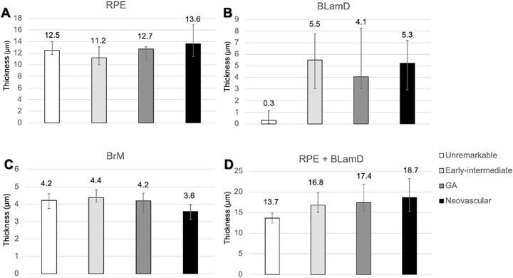
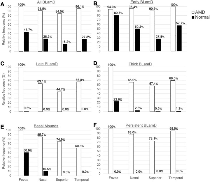
Results: In non-atrophic areas of AMD eyes, the RPE-BLamD is thick (normal = 13.7 µm, early-intermediate = 16.8 µm, GA = 17.4 µm, NV = 18.7 µm), because the BLamD is thick (normal = 0.3 µm, early-intermediate = 5.5 µm, GA = 4.1 µm, NV = 5.3 µm). RPE layer thickness is similar across these stages. Disease-associated variants of BLamD (thick, late, basal mounds) cluster subfoveally. A thick BLamD is visible on OCT as a hyporeflective split in the RPE-BL-BrM complex. BrM is thin (3.5 µm) in NV (normal = 4.2 µm, early to intermediate = 4.4 µm, and GA = 4.2 µm).

Conclusions: The RPE-BL-BrM complex is thick in AMD, driven by the accumulation and expansion of BLamD rather than expansion of either three-layer BrM, RPE-BL, or RPE. BLamD is clinically appreciable by OCT in some patients as a non-neovascular "split RPE-BL-BrM complex" or "double-layer sign." BLamD may contribute toward the formation and progression of high-risk drusen yet also exhibit protective properties.


Conflict of interest statement

Disclosure: A.A. Sura, None; L. Chen, None; J.D. Messinger, None; T.A. Swain, None; G. McGwin Jr., None; K.B. Freund, Optovue (C), Heidelberg Engineering (C), Bayer (C), Zeiss (C), Allergan (C), and Novartis (C), and Genentech/Roche (F). C.A. Curcio, Heidelberg Engineering and Genentech/Hoffman LaRoche (F), MacRegen Inc (I)

Figures

Figure 1.
Figure 1.
The layers of age-related macular degeneration include basal laminar deposit. The RPE as an epithelium rests on a basal lamina of extracellular matrix (at left). BLamD is a stereotypically thickened extracellular matrix (green, in middle and right) between the RPE plasma membrane and this basal lamina. BLamD either replaces or incorporates the RPE basal infoldings, shown as pleats at the bottom of the RPE cell body at the left). The trilaminar Bruch's membrane (BrM),,, consists of the inner collagenous layer (ICL), elastic layer (EL), and outer collagenous layer (OCL). Between the RPE basal lamina (or BLamD) and inner collagenous layer of Bruch's membrane (subRPE-BL space) accumulate soft drusen material (lipoprotein-related debris or “membranous debris”) and in some eyes type 1 MNV, of choroidal origin (double headed arrow). Soft druse material is found also in basal linear deposits (BLinD) and in basal mounds within BLamD. BLinD may be continuous with, yet distinct from drusen, which are dome-shaped. The schematic bar at the left shows the third and fourth outer retinal reflective bands of currently available commercial optical coherence tomography (OCT) with our interpretation based on recent mapping of RPE organelles, and supported by adaptive optics assisted OCT., The third band, interdigitation zone (IZ), corresponds to interleaved photoreceptor outer segments and RPE apical processes with their notable content of melanosomes. Components of the fourth band include the RPE cell body, RPE basal infoldings, RPE-BL, BLamD if present, contents of sub-RPE-BL space if present, and ICL-EL-OCL of BrM. The apical 3/4 of the RPE cell body contains approximately 700 reflective lipofuscin and melanolipofuscin granules with some melanosomes and a similar number of reflective mitochondria in basolateral 3/4 (middle half has all organelles). The unnamed hyporeflective band between IZ and RPE-BL-BrM band are currently thought to be a zone of apical processes where they depart from the cell body and lack melanosomes. An attribution to a layer of phagosomes within the RPE cell body based on selective stains was not supported by comprehensive electron microscopy. OS, outer segments of photoreceptors; RPE, retinal pigment epithelium; M, melanosome; ML, melanolipofuscin; Mt, mitochondria; RPE-BL, RPE basal lamina; BLamD, basal laminar deposit; BLinD, basal linear deposit; ChC, choriocapillaris; ChC-BL, ChC basal lamina.
Figure 2.
Figure 2.
Histopathology of basal laminar deposits (BLamD) in age-related macular degeneration (AMD). (A) Normal macula, without BLamD in a 77-year-old woman, 2000 µm nasal to the foveal center. (B) Patchy early BLamD (yellow asterisk) in a normal macula in an 88-year-old man, 600 µm nasal. (C) Early (yellow asterisk) and late (red asterisk) BLamD, along with basal linear deposit (fuchsia arrowhead), in an 85-year-old woman with early to intermediate AMD. Yellow arrowhead, ChC ghost, 400 µm temporal. (D) Thick, continuous early and late BLamD (yellow and red asterisks) in an 88-year-old man with early to intermediate AMD. A thin layer of pre-BLinD (fuchsia arrowhead) can be observed in this eye. Yellow arrowhead, ChC retraction, 290 µm nasal. (E) Persistent BLamD in the atrophic zone (yellow asterisks) of a 76-year-old woman with geographic atrophy. Müller glial processes were observed in the sub RPE-BL space. Note the continuity of BLamD across the border of atrophy, signified by the descent of the external limiting membrane (green arrowheads), 400 µm nasal. (F) Persistent BLamD overlying choroidal neovascularization (CNV) in a 91-year-old female. Both early and late (yellow and red asterisks) BLamD can be observed. Fibrovascular (fv) and fibrocellular (fc) membranes are shown on either side of the persistent BLamD, 490 µm temporal. INL, inner nuclear layer; HFL, Henle fiber layer; ONL, outer nuclear layer; IS, inner segment; OS, outer segment; RPE, retinal pigment epithelium; BrM, Bruch's membrane; ChC, choriocapillaris. Retina is detached from RPE in panels B to D.
Figure 3.
Figure 3.
Histopathology of basal mounds (BMounds) in age-related macular degeneration (AMD). (A) BMounds (red asterisk) are internal to the RPE basal lamina (yellow arrowhead) whereas drusen (d) are external to it, in a 76-year-old woman with early to intermediate AMD, 200 µm nasal. (B) BMounds (red asterisk) may appear as focal mound-like excrescences, in a 95-year-old man with early to intermediate AMD, 400 µm nasal. (C) BMounds (red asterisk) may form a semicontinuous layer of lipoprotein rich material within the BLamD. Pigmented cells of RPE origin can be observed in the retina and subretinal space (red arrowheads). Yellow arrowheads, retracted ChC and ghosts, in an 83-year-old woman with outer retinal atrophy, 1500 µm nasal. (D) BMounds can become quite large (red asterisk) and include complex features, such as internal structure (fuchsia arrowhead) and non-nucleated granule aggregates shed from RPE (orange arrowhead) in an 85-year-old woman with early to intermediate AMD, 1500 µm temporal. (E) Much like drusen, BMounds (red asterisk) may become calcified (blue arrowhead) in a 96-year-old woman with early to intermediate AMD, 2000 µm temporal. HFL, Henle fiber layer; ONL, outer nuclear layer; HFL/ONL, dyslamination of HFL and ONL; IS, inner segment; RPE, retinal pigment epithelium; BLamD, basal laminar deposit; BrM, Bruch's membrane; Ch, choroid; ChC, choriocapillaris. Green arrowhead, external limiting membrane. SDD, subretinal drusenoid deposits; Retina is detached from RPE in panels A to D.
Figure 4.
Figure 4.
Basal laminar deposit (BLamD) is ubiquitous in age-related macular degeneration (AMD). At predefined locations in the fovea and perifovea, the presence of BLamD was assayed (19–38 locations per eye) and morphologic phenotype defined. (A) Most aged eyes had at least one location with BLamD. (B) When locations are systematically sampled, BLamD is more commonly encountered in AMD. (C) BLamD phenotypes shift along different diagnostic classes of AMD. Although a feature of normal aging, early BLamD considerably accumulates in early to intermediate AMD and geographic atrophy (GA). Thick BLamD (defined as BLamD half as thick as the average RPE cell [> 6.2 µm]) and late BLamD were associated with the transition to early-intermediate AMD. Basal mounds—drusen-like accumulations of lipid within BLamD—were likewise more common in eyes with AMD. Persistent BLamD are found in areas of atrophy and represent a predominant form of BLamD in GA and neovascular AMD.
Figure 5.
Figure 5.
Thickness of the retinal pigment epithelium (RPE), basal laminar deposit (BLamD), and Bruch's membrane (BrM) in different stages of age-related macular degeneration (AMD). At predefined locations in the fovea and perifovea, RPE, BLamD, and BrM were assayed (19–38 locations per eye). Only nonzero RPE measurements were included. Measurements within the same eye were averaged, and each eye was ranked. Data represent median and interquartile range of the rank list. (A) RPE cell body thicknesses were similar between groups with more variability in advanced disease. (B) Eyes with AMD strikingly accumulate BLamD compared to eyes without disease. (C) The interquartile range of BrM thicknesses was thin in neovascular AMD (3.1–4.0), relative to normal (4.1–4.8), and early to intermediate AMD (4.0–4.6). (D) The median RPE + BLamD was 3 to 5 µm greater in AMD eyes relative to normal eyes. The thickening of the RPE-basal lamina/BrM complex in AMD is largely driven by the addition and expansion of BLamD.
Figure 6.
Figure 6.
Topography of basal laminar deposit (BLamD) phenotypes. At predefined locations in the fovea and perifovea, the presence of any BLamD, early BLamD, late BLamD, thick BLamD, basal mounds, and persistent BLamD was assayed. Fovea and regions of perifovea was defined as described in the Methods section. Thick BLamD was defined as > 6.2 µm (half the thickness of the average RPE cell body). Data are normalized to the most commonly encountered location for each diagnostic category. (A, B) In eyes with AMD, BLamD and early BLamD was widespread. (C, D) Late and thick BLamD are specific for AMD and frequently accumulate under the fovea. (E) Basal mounds cluster subfoveally, even in normal eyes.
Figure 7.
Figure 7.
Phenotypes of retinal pigment epithelium (RPE) morphology in different stages of age-related macular degeneration (AMD). At predefined locations in the fovea and perifovea, RPE phenotypes were determined using the Project MACULA RPE grading system. Data represent percentage of measurements with a given phenotype, with degree of pathology increasing down the y axis. RPE appears more dysmorphic in early-intermediate AMD than in unremarkable eyes. Eyes with geographic atrophy and neovascular AMD have few RPE cells with normal architecture. Legend: black bars, epithelial; white bars, atrophic; grey bars, nonepithelial.
Figure 8.
Figure 8.
Clinicopathologic correlation of BLamD, compared to type 1 MNV. (A) Optical coherence tomography (OCT) B-scan 16 months before patient death shows a large, central, shallow irregular RPE elevation (SIRE; red arrowheads), and a split RPE-BL-BrM complex or “double-layer sign” on the left (yellow arrowheads). Green, yellow, and red frames are magnified in panels C, E, and G, respectively. (B) Panoramic view of histology shows type 1 macular neovascularization (MNV; red arrowheads), and a thick basal laminar deposit (BLamD; yellow arrowhead), exactly corresponding with the OCT B-scan. Green, yellow, and red frames are magnified in panel D, F, and H, respectively. (C) Magnified OCT B-scan shows an area without RPE-BL elevation. The ELM is visible on the left side. The EZ band is not continuous. (D) Magnified histologic image shows a continuous RPE layer with very thin BLamD (yellow asterisk), intact ELM, and dyslaminate ONL-HFL. (E) Magnified OCT B-scan shows an area with a split RPE-BL-BrM complex. The hyper-reflective RPE band is noticeably thicker, with a thin hyporeflective band between it and Bruch's membrane. The ELM is detectable. An EZ band is not discernible. (F) Magnified histologic image shows a continuous RPE layer and short photoreceptors (PRs). A very thick layer of BLamD, including early (yellow asterisk) and late (red asterisk) forms can be observed underneath the RPE. (G) Magnified OCT B-scan shows an area with a SIRE. The RPE-BL band is thickened and the EZ band is continuous and thick. (H) Magnified histologic image shows type 1 MNV between RPE with BLamD and a thin BrM. IPL, inner plexiform layer; INL, inner nuclear layer; OPL, outer plexiform layer; HFL, Henle fiber layer; ONL, outer nuclear layer; ELM, external limiting membrane; EZ, ellipsoid zone; RPE, retinal pigment epithelium; BL, basal lamina; BrM, Bruch's membrane; IS, inner segment; OS, outer segment; ChC, choriocapillaris. Scale bar in D applies to D, F, and H.

References

    1. Flaxman SR, Bourne RRA, Resnikoff S, et al. .. Global causes of blindness and distance vision impairment 1990–2020: a systematic review and meta-analysis. Lancet Glob Health. 2017; 5: e1221–e1234. - PubMed
    1. Comparison of Age-related Macular Degeneration Treatments Trials Research Group, Maguire MG, Martin DF, et al. .. Five-year outcomes with anti-vascular endothelial growth factor treatment of neovascular age-related macular degeneration: the comparison of age-related macular degeneration treatments trials. Ophthalmology. 2016; 123: 1751–1761. - PMC - PubMed
    1. Age-Related Eye Disease Study 2 Research Group. Lutein + zeaxanthin and omega-3 fatty acids for age-related macular degeneration: the Age-Related Eye Disease Study 2 (AREDS2) randomized clinical trial. JAMA. 2013; 309: 2005–2015. - PubMed
    1. Staurenghi G, Sadda S, Chakravarthy U, Spaide RF, International Nomenclature for Optical Coherence Tomography (IN OCT) Panel. Proposed lexicon for anatomic landmarks in normal posterior segment spectral-domain optical coherence tomography: the IN*OCT consensus. Ophthalmology. 2014; 121: 1572–1578. - PubMed
    1. Gass JDM. Stereoscopic atlas of macular diseases: diagnosis and treatment. St. Louis, MO: Mosby; 1997.

Publication types