

Evaluation of Glycerylphytate Crosslinked Semi- and Interpenetrated Polymer Membranes of Hyaluronic Acid and Chitosan for Tissue Engineering
- PMID: 33187239
- PMCID: PMC7697555
- DOI: 10.3390/polym12112661
Evaluation of Glycerylphytate Crosslinked Semi- and Interpenetrated Polymer Membranes of Hyaluronic Acid and Chitosan for Tissue Engineering
Abstract
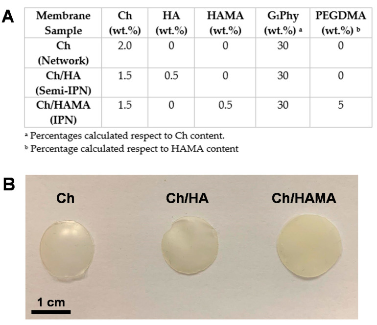
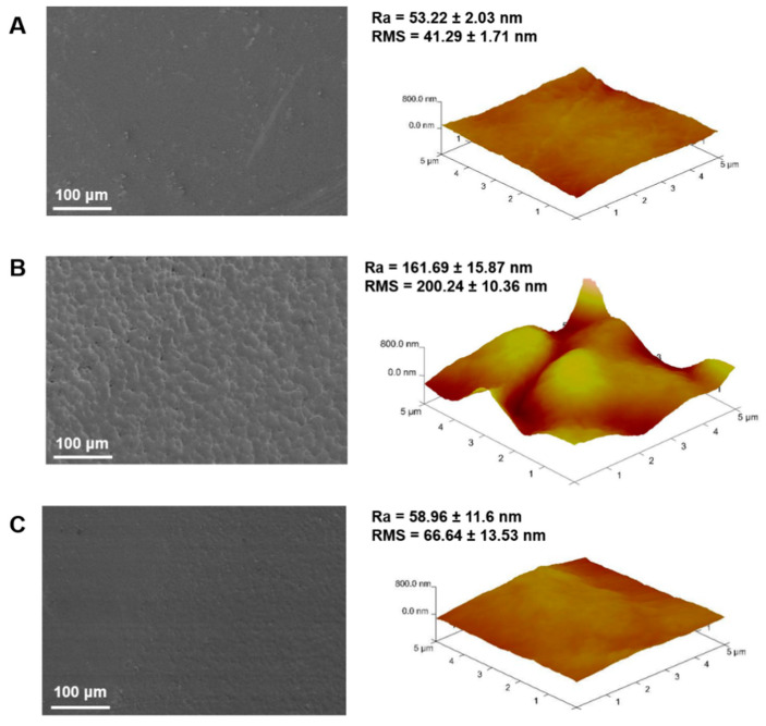
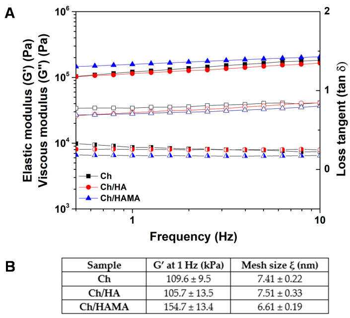
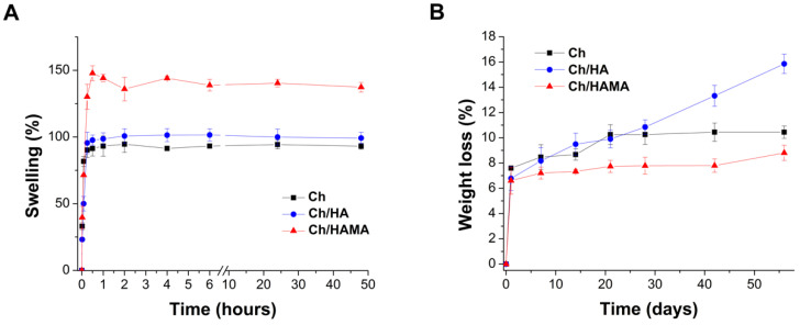
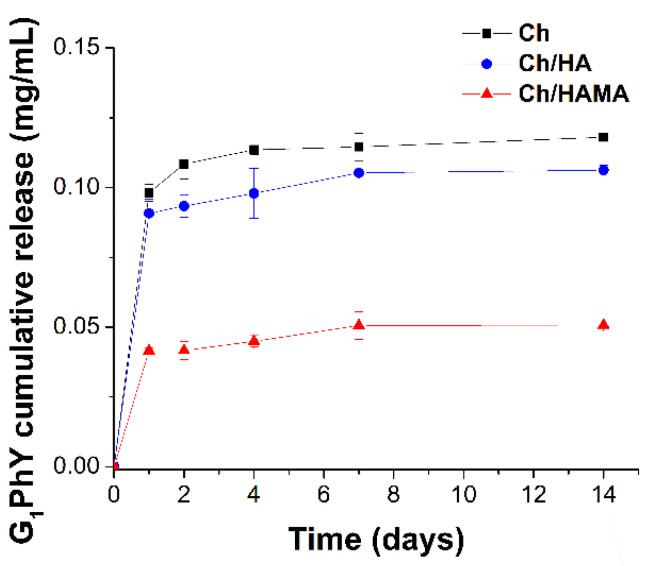
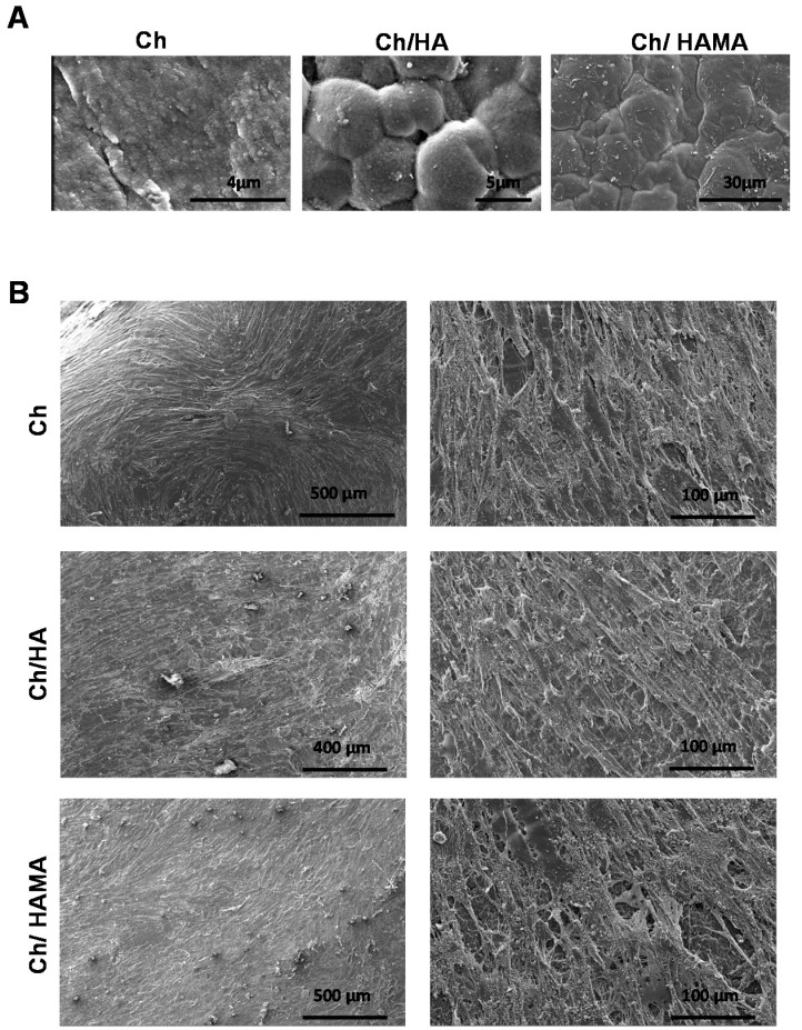
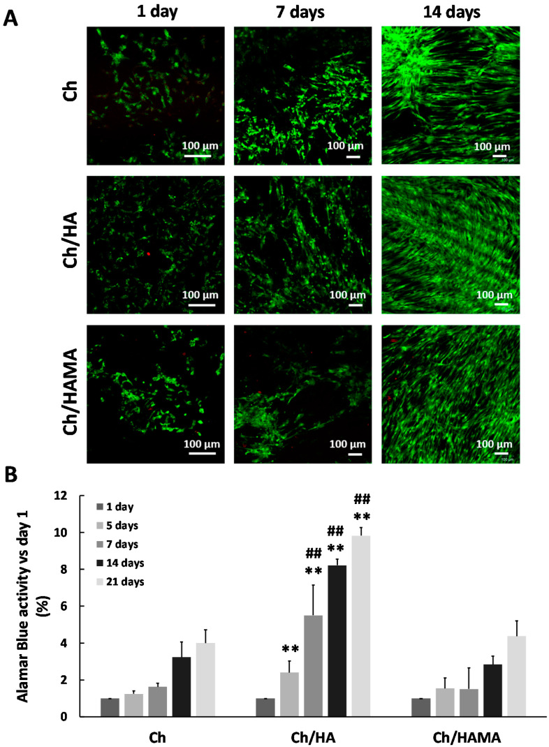
In the present study, semi- and interpenetrated polymer network (IPN) systems based on hyaluronic acid (HA) and chitosan using ionic crosslinking of chitosan with a bioactive crosslinker, glycerylphytate (G1Phy), and UV irradiation of methacrylate were developed, characterized and evaluated as potential supports for tissue engineering. Semi- and IPN systems showed significant differences between them regarding composition, morphology, and mechanical properties after physicochemical characterization. Dual crosslinking process of IPN systems enhanced HA retention and mechanical properties, providing also flatter and denser surfaces in comparison to semi-IPN membranes. The biological performance was evaluated on primary human mesenchymal stem cells (hMSCs) and the systems revealed no cytotoxic effect. The excellent biocompatibility of the systems was demonstrated by large spreading areas of hMSCs on hydrogel membrane surfaces. Cell proliferation increased over time for all the systems, being significantly enhanced in the semi-IPN, which suggested that these polymeric membranes could be proposed as an effective promoter system of tissue repair. In this sense, the developed crosslinked biomimetic and biodegradable membranes can provide a stable and amenable environment for hMSCs support and growth with potential applications in the biomedical field.
Keywords: chitosan; glycerylphytate; interpenetrated polymer network; mesenchymal stem cell; methacrylated hyaluronic acid; semi-IPN.
Conflict of interest statement
The authors declare no conflict of interest.
Figures
Similar articles
-
Glycerylphytate crosslinker as a potential osteoinductor of chitosan-based systems for guided bone regeneration.Carbohydr Polym. 2020 Aug 1;241:116269. doi: 10.1016/j.carbpol.2020.116269. Epub 2020 Apr 28. Carbohydr Polym. 2020. PMID: 32507162
-
Glycerylphytate as an ionic crosslinker for 3D printing of multi-layered scaffolds with improved shape fidelity and biological features.Biomater Sci. 2019 Dec 17;8(1):506-516. doi: 10.1039/c9bm01271k. Biomater Sci. 2019. PMID: 31764919
-
Bioadhesive functional hydrogels: Controlled release of catechol species with antioxidant and antiinflammatory behavior.Mater Sci Eng C Mater Biol Appl. 2019 Dec;105:110040. doi: 10.1016/j.msec.2019.110040. Epub 2019 Jul 31. Mater Sci Eng C Mater Biol Appl. 2019. PMID: 31546368
-
Weaving the next generation of (bio)materials: Semi-interpenetrated and interpenetrated polymeric networks for biomedical applications.Adv Colloid Interface Sci. 2023 Oct 21;321:103026. doi: 10.1016/j.cis.2023.103026. Online ahead of print. Adv Colloid Interface Sci. 2023. PMID: 39491440 Review.
-
Interpenetrating Polymer Networks polysaccharide hydrogels for drug delivery and tissue engineering.Adv Drug Deliv Rev. 2013 Aug;65(9):1172-87. doi: 10.1016/j.addr.2013.04.002. Epub 2013 Apr 17. Adv Drug Deliv Rev. 2013. PMID: 23603210 Review.
Cited by
-
Development of Covalent Chitosan-Polyethylenimine Derivatives as Gene Delivery Vehicle: Synthesis, Characterization, and Evaluation.Int J Mol Sci. 2021 Apr 7;22(8):3828. doi: 10.3390/ijms22083828. Int J Mol Sci. 2021. PMID: 33917124 Free PMC article.
-
Self-Associating Polymers Chitosan and Hyaluronan for Constructing Composite Membranes as Skin-Wound Dressings Carrying Therapeutics.Molecules. 2021 Apr 26;26(9):2535. doi: 10.3390/molecules26092535. Molecules. 2021. PMID: 33926140 Free PMC article. Review.
-
Chitosan Functionalization: Covalent and Non-Covalent Interactions and Their Characterization.Polymers (Basel). 2021 Nov 26;13(23):4118. doi: 10.3390/polym13234118. Polymers (Basel). 2021. PMID: 34883621 Free PMC article. Review.
References
-
- Gilarska A., Lewandowska-Lancucka J., Horak W., Nowakowska M. Collagen/chitosan/hyaluronic acid-based injectable hydrogels for tissue engineering applications—Design, physicochemical and biological characterization. Colloids Surf. B Biointerfaces. 2018;170:152–162. doi: 10.1016/j.colsurfb.2018.06.004. - DOI - PubMed
-
- Nair S., Remya N.S., Remya S., Nair P.D. A biodegradable in situ injectable hydrogel based on chitosan and oxidized hyaluronic acid for tissue engineering applications. Carbohydr. Polym. 2011;85:838–844. doi: 10.1016/j.carbpol.2011.04.004. - DOI
-
- Suo H., Zhang D., Yin J., Qian J., Wu Z.L., Fu J. Interpenetrating polymer network hydrogels composed of chitosan and photocrosslinkable gelatin with enhanced mechanical properties for tissue engineering. Mater. Sci. Eng. C Mater. Biol. Appl. 2018;92:612–620. doi: 10.1016/j.msec.2018.07.016. - DOI - PubMed
