. 2020 Nov 11;9(11):1113.
doi: 10.3390/antiox9111113.

Modulation of the Neuroprotective and Anti-inflammatory Activities of the Flavonol Fisetin by the Transition Metals Iron and Copper

Affiliations

Modulation of the Neuroprotective and Anti-inflammatory Activities of the Flavonol Fisetin by the Transition Metals Iron and Copper

Pamela Maher. Antioxidants (Basel). .

Abstract

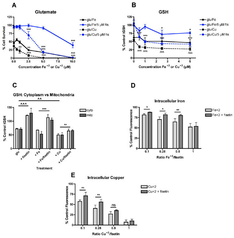
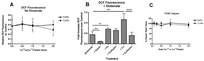
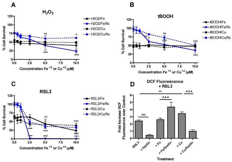
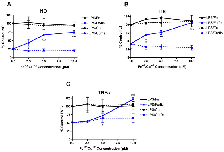
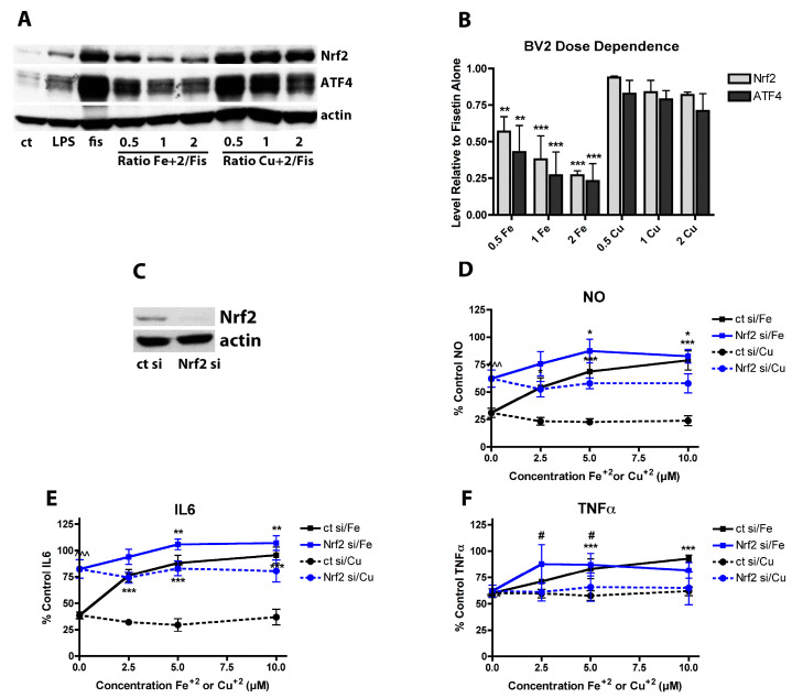
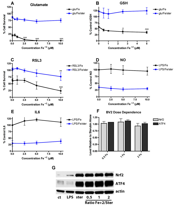
Alterations occur in the homeostasis of the transition metals iron (Fe2+) and copper (Cu2+) during aging and these are further amplified in neurodegenerative diseases, including Alzheimer's disease (AD). These observations suggest that the most effective drug candidates for AD might be those that can reduce these alterations. The flavonoid fisetin has both neuroprotective and anti-inflammatory activity both in vitro and in vivo and can bind both iron and copper suggesting that its chelating activity might play a role in its beneficial effects. To test this idea, the effects of iron and copper on both the neuroprotective and anti-inflammatory activities of fisetin were examined. It is shown that while fisetin can reduce the potentiation of cell death by iron and copper in response to treatments that lower glutathione levels, it is much less effective when the metals are combined with other inducers of oxidative stress. In addition, iron but not copper reduces the anti-inflammatory effects of fisetin in a dose-dependent manner. These effects correlate with the ability of iron but not copper to block the induction of the antioxidant transcription factor, Nrf2, by fisetin. In contrast, although the flavanone sterubin also binds iron, the metal has no effect on sterubin's ability to induce Nrf2 or protect cells from toxic or pro-inflammatory insults. Together, these results suggest that while iron and copper binding could contribute to the beneficial effects of neuroprotective compounds in the context of neurodegenerative diseases, the consequences of this binding need to be fully examined for each compound.

Keywords: Alzheimer’s disease; Nrf2; ferroptosis; glutathione; oxidative stress; oxytosis; sterubin.


Conflict of interest statement

The author declares no conflict of interest.

Figures

Figure 1
Figure 1
FeCl2 and CuCl2 modulate the effects of fisetin on survival and GSH. (A) Fisetin reduces the potentiation of glutamate toxicity by FeCl2 and CuCl2. HT22 cells were treated with 2.5 mM glutamate in the presence of 5 µM fisetin and/or FeCl2 or CuCl2 at the indicated concentrations. Cell survival was measured after 24 h with the MTT assay. The experiments were done in quadruplicate and the results are the average of 5–6 independent experiments. (B) Fisetin reduces the potentiation of total GSH loss by FeCl2 and CuCl2. HT22 cells were treated with 2.5 mM glutamate in the presence of 5 µM fisetin and/or FeCl2 or CuCl2 at the indicated concentrations. Cells were harvested after 24 h and total GSH was measured. The results are the average of 5-6 independent experiments. * indicates p < 0.05 and *** indicates p < 0.001 relative to fisetin + glutamate alone and ^ indicates p < 0.05, ^^ indicates p < 0.01 and ^^^ indicates p < 0.001 relative to glutamate alone. (C) Fisetin similarly reduces the potentiation of total GSH loss by FeCl2 and CuCl2 in both cytoplasm and mitochondria. HT22 cells were treated with 2.5 mM glutamate in the presence of 10 µM fisetin and/or 5 µM FeCl2 or CuCl2 (0.5:1, metal:fisetin). Cells were harvested after 24 h, separated into cytoplasmic and mitochondrial fractions and total GSH was measured. The results are the average of 4–6 independent experiments. ** indicates p < 0.01 and *** indicates p < 0.001 relative to fisetin + glutamate alone. ^^ indicates p < 0.01 and ^^^ indicates p < 0.001 relative to glutamate alone. (D) Fisetin reduces free intracellular Fe2+ levels at Fe2+:fisetin ratios of 0.5:1 and below. Free intracellular Fe2+ was measured using the dye PhenGreen SK after 3 h of treatment with 10 µM fisetin and the indicated concentrations of FeCl2. (E) Fisetin reduces free intracellular Cu2+ levels at Cu2+:fisetin ratios of 0.25:1 and below. Free intracellular Cu2+ was measured using the dye PhenGreen FL after 3 h of treatment with 10 µM fisetin and the indicated concentrations of CuCl2. The experiments were done in sextuplicate and the results are the average of 4–6 independent experiments. * indicates p < 0.05 and ** indicates p < 0.01 relative to the metal alone.
Figure 2
Figure 2
Effects of FeCl2 and CuCl2 on fisetin-induced increases in the transcription factors Nrf2 and ATF4. (A) Representative Western blots for Nrf2, ATF4 and actin of nuclear extracts from HT22 cells treated for 4 h with 5 µM fisetin either alone (fis) or in the presence of increasing concentrations of FeCl2 or CuCl2. (B) Quantitation of results from four independent experiments identical to (A). (C) Representative Western blots for Nrf2, ATF4 and actin of nuclear extracts from HT22 cells treated for 4 h with 5 µM fisetin, 1 µM carnosol, 1 µM celastrol or 5 µM sulforaphane either alone (-) or in the presence of a 2 fold molar excess of FeCl2 or CuCl2. (D) Quantitation of results from three independent experiments identical to (C). Since sulforaphane did not induce ATF4, no results for this are shown. (E) Representative Western blots for Nrf2, ATF4 and actin of nuclear extracts from HT22 cells treated for 4 h with 5 µM fisetin, 5 µM quercetin or 5 µM sterubin either alone (-) or in the presence of 10 µM FeCl2 or CuCl2. (F) Quantitation of results from four independent experiments identical to (E). In (B,D), * indicates p < 0.05, ** indicates p < 0.01 and *** indicates p < 0.001 relative to fisetin alone. In (F), * indicates p < 0.05 and *** indicates p < 0.001 relative to compound alone.
Figure 3
Figure 3
Effects of FeCl2 and CuCl2 on fisetin-mediated reductions in ROS levels. (A) HT22 cells were treated with 10 µM fisetin and increasing concentrations of FeCl2 or CuCl2. After 7 hr, ROS levels were determined using CM-H2DCFDA as described in Materials and Methods. The treatments were done in sextuplicate and the results are the average of 4 independent experiments. (B) Cells were treated with 5 mM glutamate alone or in the presence of 10 µM fisetin and/or 10 µM of FeCl2 or CuCl2. After 7 h, ROS levels were determined using CM-H2DCFDA as described in Materials and Methods. The treatments were done in sextuplicate and the results are the average of 4 independent experiments. ** p < 0.01, *** p < 0.001 relative to glutamate alone. ^^^ p < 0.001 relative to glutamate + CuCl2 alone. (C) TEAC values were determined for fisetin in the presence of increasing concentrations of FeCl2 or CuCl2 as described in Materials and Methods.
Figure 4
Figure 4
FeCl2 and CuCl2 modulate the effects of fisetin on protection from multiple toxicities. FeCl2 and CuCl2 reduce fisetin-mediated protection against hydrogen peroxide (H2O2) (A) or t-butyl peroxide (tBOOH) (B) toxicity. HT22 cells were treated with 0.5 mM H2O2 or 2.5 µM tBOOH in the presence of 10 µM fisetin and/or FeCl2 or CuCl2 at the indicated concentrations. Cell survival was measured after 24 h with the MTT assay. The experiments were done in quadruplicate and the results are the average of 4 independent experiments. (C) FeCl2 and CuCl2 differentially affect fisetin-mediated protection against RSL3 toxicity. HT22 cells were treated with 100 nM RSL3 in the presence of 5 µM fisetin and/or FeCl2 or CuCl2 at the indicated concentrations. Cell survival was measured after 24 h with the MTT assay. The experiments were done in quadruplicate and the results are the average of 5 independent experiments. (AC), * indicates p < 0.05, ** indicates p < 0.01 and *** indicates p < 0.001 relative to fisetin + toxin alone. (C), ^^ indicates p < 0.01 and ^^^ indicates p < 0.001 relative to RSL3 alone for both FeCl2 and CuCl2. (D) Cells were treated with 250 nM RSL3 alone or in the presence of 10 µM fisetin and 5 µM FeCl2 or CuCl2. After 4 h, ROS levels were determined using CM-H2DCFDA as described in Materials and Methods. The treatments were done in sextuplicate and the results are the average of 4 independent experiments. ** p < 0.01, *** p < 0.001 relative to RSL3 alone. ^^^ p < 0.001 relative to RSL3 + FeCl2 or CuCl2 alone.
Figure 5
Figure 5
FeCl2 and CuCl2 differentially modulate the anti-inflammatory effects of fisetin. Dose dependent effects of FeCl2 and CuCl2 on the inhibition of NO and pro-inflammatory cytokine production by fisetin in LPS-treated BV-2 microglial cells. BV-2 cells were treated overnight with 25 ng/mL LPS in the presence of 5 µM fisetin alone or with the addition of the indicated concentrations of FeCl2 or CuCl2. Cell culture supernatants were cleared and assayed for NO by the Griess assay or pro-inflammatory cytokines using specific ELISAs. Results are presented as the percent (%) of the value obtained with LPS alone which was set at 100%. (A) NO, (B) IL6, (C) TNFα. The results represent the average of 3–4 independent experiments. * indicates p < 0.05, ** indicates p < 0.01 and *** indicates p < 0.001 relative to fisetin + LPS alone.
Figure 6
Figure 6
Nrf2 plays a key role in the anti-inflammatory effects of fisetin. (A) Representative Western blots for Nrf2, ATF4 and actin in nuclear extracts from BV-2 cells treated with 25 ng/mL LPS alone or in the presence of 5 µM fisetin ± the indicated concentrations of FeCl2 or CuCl2 for 4 h. (B) Quantitation of results from four independent experiments identical to (A). ** indicates p < 0.01 and *** indicates p < 0.001 relative to fisetin alone. (C) Representative Western blot of BV-2 cells treated for 48 h with control siRNA (ct si) or Nrf2 siRNA (Nrf2 si). (DF) BV2 cells transfected with control siRNA (ct si) or Nrf2 siRNA (Nrf2 si) were treated overnight with 25 ng/mL LPS and 5 µM fisetin and/or the indicated concentrations of FeCl2 or CuCl2. Cell culture supernatants were cleared and assayed for NO by the Griess assay or pro-inflammatory cytokines using specific ELISAs. Results are presented as the percent (%) of the value obtained with LPS alone which was set at 100%. NO (D); IL6 (E) or TNFα (F). Results in (DF) are the average of three independent experiments. * indicates p < 0.05, ** indicates p < 0.01 and *** indicates p < 0.001 relative to fisetin + LPS alone. # indicates p < 0.05 relative to Nrf2 siRNA with fisetin and LPS only.
Figure 7
Figure 7
FeCl2 does not interfere with the activities of sterubin. (A) FeCl2 does not alter the protective effect of sterubin against glutamate toxicity. HT22 cells were treated with 2.5 mM glutamate in the presence of 5 µM sterubin and/or FeCl2 at the indicated concentrations. Cell survival was measured after 24 h with the MTT assay. The experiments were done in quadruplicate and the results are the average of 4 independent experiments. (B) FeCl2 does not affect the maintenance of GSH levels by sterubin. HT22 cells were treated with 2.5 mM glutamate in the presence of 5 µM sterubin and/or FeCl2 at the indicated concentrations. Cells were harvested after 24 h and total GSH was measured. The results are the average of 5–6 independent experiments. (C) FeCl2 only slightly impairs the protective effect of sterubin against RSL3 toxicity. HT22 cells were treated with 100 nM RSL3 in the presence of 5 µM sterubin and/or FeCl2 at the indicated concentrations. Cell survival was measured after 24 h with the MTT assay. The experiments were done in quadruplicate and the results are the average of 4 independent experiments. (DE) FeCl2 does not reduce the anti-inflammatory effects of sterubin. BV-2 cells were treated overnight with 25 ng/mL LPS in the presence of 5 µM sterubin alone or with the addition of the indicated concentrations of FeCl2. Cell culture supernatants were cleared and assayed for NO by the Griess assay or IL-6 using a specific ELISA. Results are presented as the percent (%) of the value obtained with LPS alone which was set at 100%. ** indicates p < 0.01 relative to sterubin + RSL3 alone. ^ indicates p < 0.05 and ^^^ indicates p < 0.001 relative to insult alone. (F) Quantitation of results from four independent experiments identical to (G). (G) Representative Western blots for Nrf2, ATF4 and actin of nuclear extracts from BV-2 cells treated with 25 ng/mL LPS alone or in the presence of 5 µM sterubin ± the indicated concentrations of FeCl2 or CuCl2 for 4 h.

Similar articles

Cited by

References

    1. Maher P. Preventing and treating neurological disorders with the flavonol fisetin. Brain Plast. 2020 doi: 10.3233/BPL-200104. in press. - DOI - PMC - PubMed
    1. Ishige K., Schubert D., Sagara Y. Flavonoids protect neuronal cells from oxidative stress by three distinct mechanisms. Free Radic. Biol. Med. 2001;30:433–446. doi: 10.1016/S0891-5849(00)00498-6. - DOI - PubMed
    1. Ehren J.L., Maher P. Concurrent regulation of the transcription factors Nrf2 and ATF4 mediates the enhancement of glutathione levels by the flavonoid fisetin. Biochem. Pharmacol. 2013;85:1816–1826. doi: 10.1016/j.bcp.2013.04.010. - DOI - PubMed
    1. Currais A., Prior M., Dargusch R., Armando A., Ehren J., Schubert D., Quehenberger O., Maher P. Modulation of p25 and inflammatory pathways by fisetin maintains cognitive function in Alzheimer’s disease transgenic mice. Aging Cell. 2014;13:379–390. doi: 10.1111/acel.12185. - DOI - PMC - PubMed
    1. Currais A., Farrokhi C., Dargusch R., Armando A., Quehenberger O., Schubert D., Maher P. Fisetin reduces the impact of aging on behavior and physiology in the rapidly aging SAMP8 mouse. J. Gerentol. A Biol. Sci. Med. Sci. 2018;73:299–307. doi: 10.1093/gerona/glx104. - DOI - PMC - PubMed

LinkOut - more resources