Review
. 2020 Nov 11;25(22):5256.
doi: 10.3390/molecules25225256.

Advances in Optical Detection of Human-Associated Pathogenic Bacteria

Affiliations
Review

Advances in Optical Detection of Human-Associated Pathogenic Bacteria

Andrea Locke et al. Molecules. .

Abstract

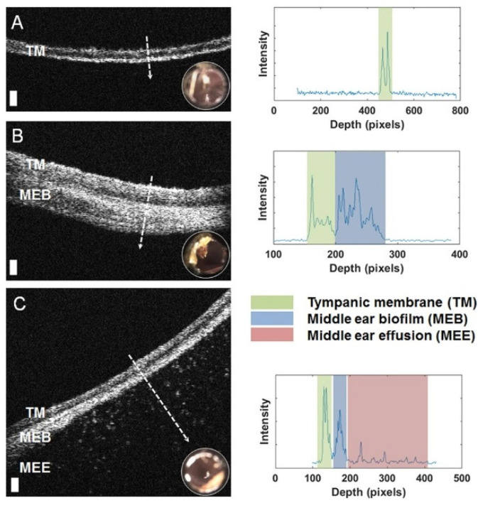
Bacterial infection is a global burden that results in numerous hospital visits and deaths annually. The rise of multi-drug resistant bacteria has dramatically increased this burden. Therefore, there is a clinical need to detect and identify bacteria rapidly and accurately in their native state or a culture-free environment. Current diagnostic techniques lack speed and effectiveness in detecting bacteria that are culture-negative, as well as options for in vivo detection. The optical detection of bacteria offers the potential to overcome these obstacles by providing various platforms that can detect bacteria rapidly, with minimum sample preparation, and, in some cases, culture-free directly from patient fluids or even in vivo. These modalities include infrared, Raman, and fluorescence spectroscopy, along with optical coherence tomography, interference, polarization, and laser speckle. However, these techniques are not without their own set of limitations. This review summarizes the strengths and weaknesses of utilizing each of these optical tools for rapid bacteria detection and identification.

Keywords: OCT; Raman; bacterial infection; fluorescence; infrared; optical detection.


Conflict of interest statement

The authors declare no conflict of interest.

Figures

Figure 1
Figure 1
Vibrational spectra of Streptomyces pseudovenezuela (a) IR absorption spectrum, (b) micro-Raman spectrum with an excitation wavelength of 532 nm, and (c) UV-resonance Raman spectrum with an excitation wavelength of 244 nm. Reprinted with permission from [29]. Copyright © 2020, published by the International Society for Advancement of Cytometry.
Figure 2
Figure 2
Typical unprocessed SERS spectra showing examples from UTI isolates. Each spectrum took 10 s to collect. (A) Enterococcus spp.; (B) P. mirabilis; (C) E. coli; (D) K. pneumoniae; (E) K. oxytoca; (F) C. freundii. The maximum Raman photon count for each spectrum is given on the left. Counts on the order of thousands and tens of thousands clearly indicate that these spectra result from the SERS process rather than normal Raman scattering. Reprinted with permission from [109], Copyright © 2004 American Chemical Society.
Figure 3
Figure 3
Intramucosal bacteria in human colon identified at confocal laser endomicroscopy and fluorescence in situ hybridization (FISH). (A) Fluorescent intramucosal bacteria within the lamina propria can readily be identified using fluorescein-aided endomicroscopy. Single crypts are shown with their characteristic round appearance (blue arrow). Single bacteria and clustered bacteria (orange arrow) can be identified within the lamina propria between two crypts (pericryptal space). (B) FISH testing confirmed the presence of intramucosal bacteria due to the bright red fluorescence. The nuclei and RNA are shown in blue. Reprinted with permission from [133]. Copyright © 2020, published by BMJ Publishing Group Ltd. and the British Society of Gastroenterology.
Figure 4
Figure 4
Selective targeting and imaging of single bacteria on a smartphone. (A) Photographs of a smartphone microscope displaying images of fluorescently labeled Cronobacter spp. bacteria. (B) 3D illustration of the same optomechanical unit that is mounted on the smartphone in (A). (C) Schematic illustration of the bacterial detection procedure. Bacteria from the contaminated sample are fixed on 22 × 50 mm2 glass slides, and the bacterial membrane is permeabilized in order for the peptide nucleic acid (PNA) probe to enter the bacteria. An Alexa Fluor 488 dye is chemically linked to the PNA probe that, in turn, is designed to bind specifically to certain regions of the ribosomal RNA (rRNA) of the bacteria. After washing away unbound probes, only the targeted bacteria remain fluorescent and can be imaged using the smartphone-based microscope shown in (A). Reprinted with permission from [168]. Published by The Royal Society of Chemistry.
Figure 5
Figure 5
Left: Representative optical coherence tomography (OCT) cross-sectional (B-scan) images and A-line profiles. (A) OCT and digital otoscopy (inset) data from a normal ear. (B) Data from an ear with a middle ear biofilm (MEB). The A-line profile shows additional scattering behind the tympanic membrane (TM). (C) Subject with middle ear fluid (MEF) and an MEB. Right: The scattering profile shows three distinct regions in the scan. White dashed lines denote the location of the A-line scan within the OCT B-scan. Scale bars represent 100 micrometers in depth. Reprinted with permission from [178] Copyright © 2020, published by Springer Nature.
Figure 6
Figure 6
(a) Relative intensity change of the interference signal upon exposure to Lactobacillus acidophilus bacterial suspensions (107 cells per mL). First, a baseline was established in buffer solution After incubation with bacteria suspension, the biosensor was extensively washed before continued signal readout Note: the intensity values were normalized to the initial average intensity, marked as intensity. (b) Microscope image taken immediately after the biosensing experiment depicts L. acidophilus cells captured onto the aptamer-modified PSiO2. Reprinted with permission from [183]. Published by The Royal Society of Chemistry.
Figure 7
Figure 7
Schematic representation of biofunctionalization on Mach-Zehnder interferometer (MZI). Here, Sl is the length of the sensor area, L is the length of the sensor arm, d is the distance between the sensor and reference arms, and θ is the opening angle of the Y-divisor for angular Y-junctions, whereas R is the radius of curvature of the Y-divisor for S-bend Y-junctions. Reproduced with permission from [185]. Copyright © 2020, published by Springer Nature.
Figure 8
Figure 8
Calculated backscattering Mueller matrix images of five different bacterial colonies: (a) Escherichia coli, (b) Lactobacillus rhamnosus, (c) Rhodococcus erythropolis, (d) Staphylococcus aureus, and (e) bacteria-free Luria broth agar media. Scale bar is 2 mm. Reprinted with permission from [187]. Copyright © 2020, published by Springer Nature.
Figure 9
Figure 9
Optical scatter patterns of Listeria species and image analysis. (A) Colony scatter patterns were captured using bacterial rapid detection using optical light-scattering technology (BARDOT) at different incubation times for eight Listeria species on brain heart infusion agar plates. The rectangular selection with broken line depicts the optimal incubation time (22 h) that yielded differentiating scatter images when the colony size was 1.1 ± 0.2 mm diameter. (B) Principal component analysis of the eight Listeria species used to build the scatter image library. Blue oval selections indicate grouping of the Listeria species. (C) Principal component analysis of Listeria monocytogenes and Listeria innocua colony scatter images that were used to build a two-species scatter image library. The blue oval selections indicate the grouping of each Listeria species. Reprinted with permission from [191]. Published by Multidisciplinary Digital Publishing Institute.

References

    1. Smith K.F., Goldberg M., Rosenthal S., Carlson L., Chen J., Chen C., Ramachandran S. Global rise in human infectious disease outbreaks. J. Royal Soc. Interface. 2014;11:20140950. doi: 10.1098/rsif.2014.0950. - DOI - PMC - PubMed
    1. Centers for Disease Control and Prevention About Antibiotic Resistance. [(accessed on 3 July 2019)];2013 Available online: https://www.cdc.gov/drugresistance/about.html.
    1. Maragakis L.L., Perencevich E.N., Cosgrove S.E. Clinical and economic burden of antimicrobial resistance. Expert Rev. Anti-Infect. Ther. 2008;6:751–763. doi: 10.1586/14787210.6.5.751. - DOI - PubMed
    1. Grant S.S., Hung D.T. Persistent bacterial infections, antibiotic tolerance, and the oxidative stress response. Virulence. 2013;4:273–283. doi: 10.4161/viru.23987. - DOI - PMC - PubMed
    1. Sheikhzadeh E., CHamsaz M., Turner A., Jager E., Beni V. Label-free impedimetric biosensor for Salmonella Typhimurium detection based on poly [pyrrole-co-3-carboxyl-pyrrole] copolymer supported aptamer. Biosens. Bioelectr. 2016;80:194–200. doi: 10.1016/j.bios.2016.01.057. - DOI - PubMed