. 2020 Nov 11;21(22):8498.
doi: 10.3390/ijms21228498.

Characterization of Mesenchymal Stem Cell Differentiation within Miniaturized 3D Scaffolds through Advanced Microscopy Techniques

Affiliations

Characterization of Mesenchymal Stem Cell Differentiation within Miniaturized 3D Scaffolds through Advanced Microscopy Techniques

Valentina Parodi et al. Int J Mol Sci. .

Abstract

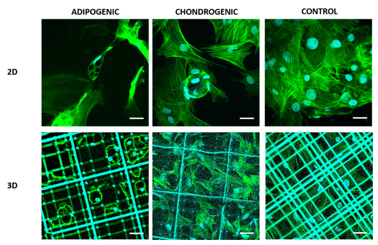
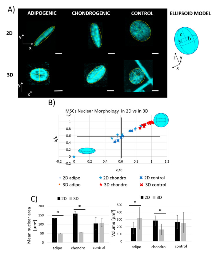
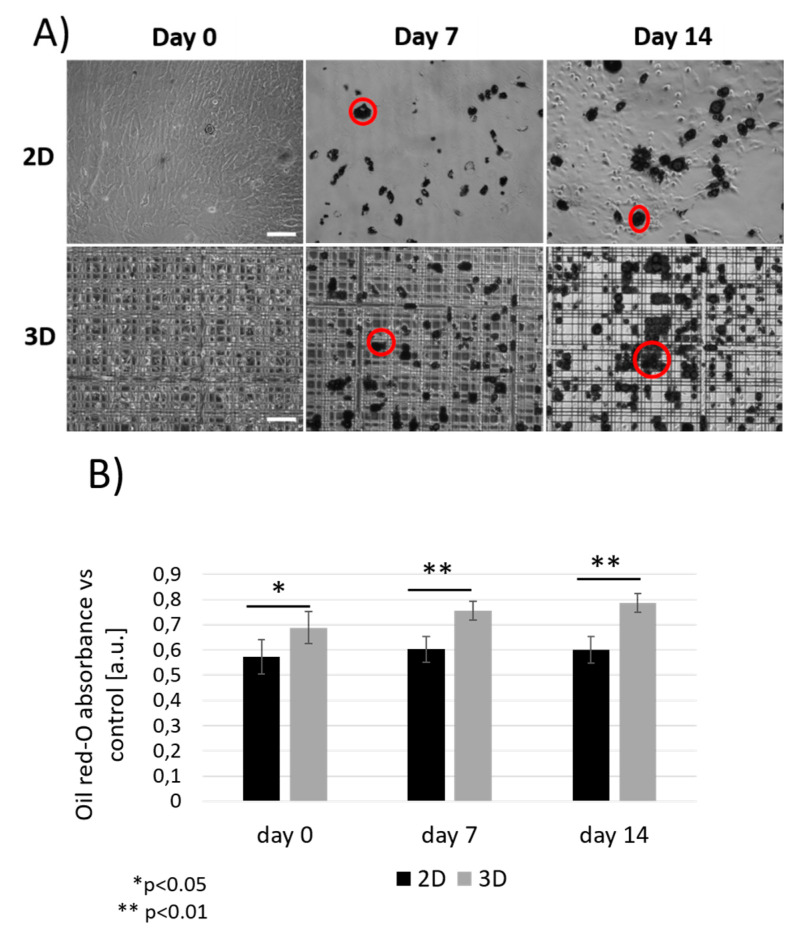
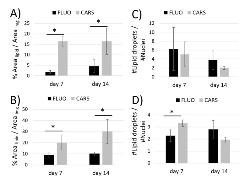
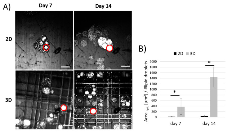
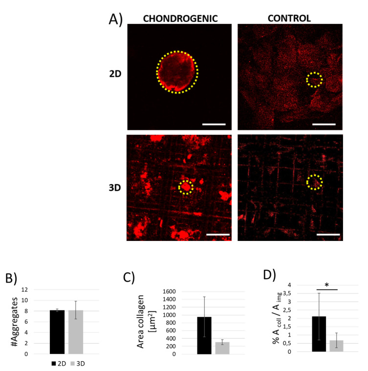
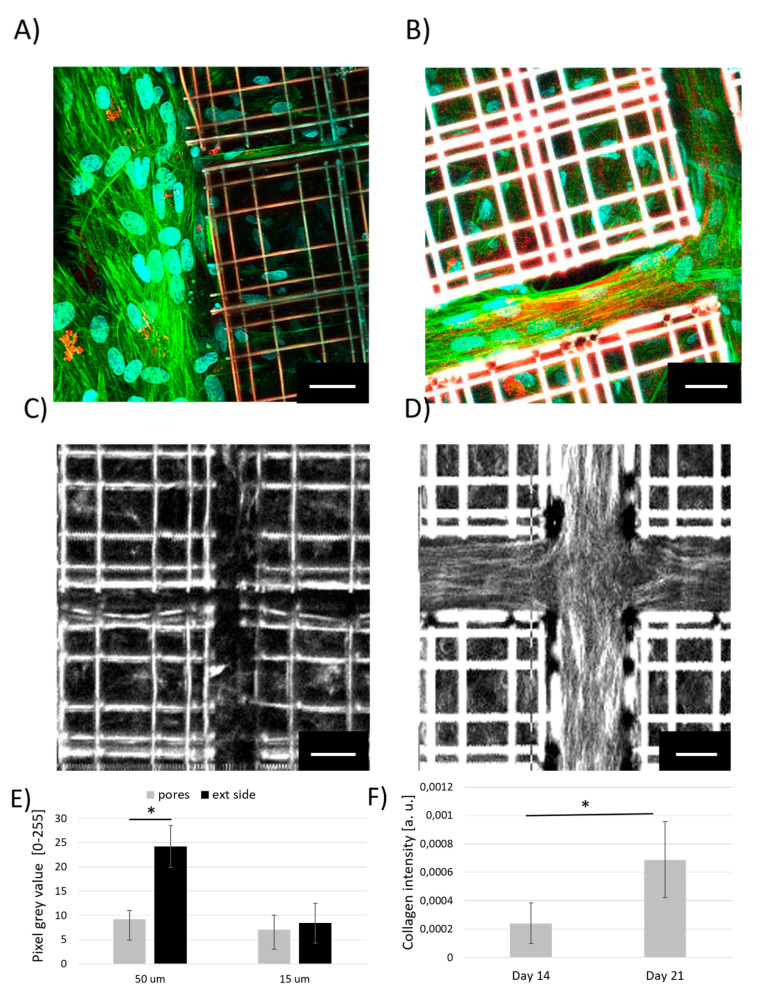
Three-dimensional culture systems and suitable substrates topographies demonstrated to drive stem cell fate in vitro by mechanical conditioning. For example, the Nichoid 3D scaffold remodels stem cells and shapes nuclei, thus promoting stem cell expansion and stemness maintenance. However, the mechanisms involved in force transmission and in biochemical signaling at the basis of fate determination are not yet clear. Among the available investigation systems, confocal fluorescence microscopy using fluorescent dyes enables the observation of cell function and shape at the subcellular scale in vital and fixed conditions. Contrarily, nonlinear optical microscopy techniques, which exploit multi-photon processes, allow to study cell behavior in vital and unlabeled conditions. We apply confocal fluorescence microscopy, coherent anti-Stokes Raman scattering (CARS), and second harmonic generation (SHG) microscopy to characterize the phenotypic expression of mesenchymal stem cells (MSCs) towards adipogenic and chondrogenic differentiation inside Nichoid scaffolds, in terms of nuclear morphology and specific phenotypic products, by comparing these techniques. We demonstrate that the Nichoid maintains a rounded nuclei during expansion and differentiation, promoting MSCs adipogenic differentiation while inhibiting chondrogenesis. We show that CARS and SHG techniques are suitable for specific estimation of the lipid and collagenous content, thus overcoming the limitations of using unspecific fluorescent probes.

Keywords: 3D culture; CARS; SHG; nonlinear microscopy; stem cell differentiation.


Conflict of interest statement

M.T.R., G.C. and R.O. are co-founders of a university spin-off company, MOAB S.r.l., and hold shares.

Figures

Figure 1
Figure 1
Immunofluorescence images of mesenchymal stem cells (MSCs) at 21 days of culture on flat substrates (2D) and on Nichoid scaffolds (3D). In green F-actin stained by phalloidin-FITC and in cyan, cell nuclei marked with Hoechst 33342. Scale bars 30 μm.
Figure 2
Figure 2
MSCs nuclear morphology assessment inside Nichoids (3D) and on controls (2D) after 21 days of culture with adipogenic, chondrogenic, and basal media. (A) Fluorescence images of single cell nuclei stained with Hoechst 33342 obtained by confocal fluorescence microscopy in the different culture conditions and schematic representation of the ellipsoid model of the MSCs nucleus with evidenced semi-axes: a, major, b, minor and c, vertical. Scale bars 5 μm. (B) Scatter plot representing the nuclei of differentiated and control cells on 3D Nichoids and on flat glass substrates, in order to compare their nuclear morphologies by means of the relationship on the ratio between the minor (b) axis with respect to the vertical (c) semi-axis and the ratio between the major (a) with respect to the vertical (c) semi-axis, respectively. These calculations allowed to investigate the effect of the Nichoid on the nuclear shape in terms of nuclear flatness and elongation. All the parameters considered were measured by Fiji-ImageJ Measure tool for ellipsoid fit, as described in Materials and Methods. (C) On the left: quantification of the mean nuclear area of nuclei measured using the maximum projection of all the acquired nuclear planes; on the right, mean nuclear volume, calculated as described in Materials and Methods. * p < 0.05.
Figure 3
Figure 3
Oil red-O staining of MSCs cultured with adipogenic differentiation media both inside Nichoids (3D) and on glass substrates (2D). (A) Brightfield images of differentiated cells taken after 24 h, 7 and 14 days from the beginning of the experiment, in which lipids appeared in dense dark aggregates. Examples of lipid vesicles are highlighted by red circles. Scale bar 100 μm. (B) Oil red-O absorbance measured at 490 nm at each time point for both 2D and 3D culture configurations. Data were normalized with respect to values from pure oil red-O and undifferentiated controls. ** p < 0.01, * p < 0.05 from pair wise comparison.
Figure 4
Figure 4
Left: day 14 of MSCs adipogenic differentiation fluorescently labeled, with Hoechst 33342 for nuclei, cyan, and DiO-lipophilic dye, green, for lipids observed through confocal fluorescence microscopy, 512 × 512 pixels. Right: vital and label free imaging obtained through coherent anti-Stokes Raman scattering (CARS) microscopy: 200 × 200 pixels, measured at 2845 cm−1. Lipid vesicles are indicated by white arrows. Scale bars 25 μm.
Figure 5
Figure 5
In the graphs, a comparison between confocal fluorescence microscopy and CARS microscopy to assess the lipid amount by calculating the ratio between the area occupied by lipids and the area of the image (A,B) and the ratio between the number of nuclei and the number of droplets in both culture configurations (C,D). * p < 0.05.
Figure 6
Figure 6
(A) Day 7 and 14 analysis of MSCs adipogenic differentiation obtained by vital and-label free CARS microscopy: 140 × 140 μm2 at 2845 cm−1. Examples of lipid vesicles are indicated by red circles. Scale bars 20 μm. (B) Differentiation efficiency represented by the ratio between the total area occupied by lipid droplets in CARS images (140 × 140 μm2) and the number of droplets counted in CARS images at day 7 and 14 of differentiation. * p < 0.05.
Figure 7
Figure 7
Indirect collagen measurements of MSCs exposed to chondrogenic and basal media at 21 days from the seeding. (A) Immunofluorescence images showing in red the signal corresponding to collagen type-I stained with anti-collagen I conjugated to Alexa Fluor 647. Examples of collagen bundles synthetized by MSCs both on glass substrates (2D) and on Nichoid scaffolds (3D), are indicated by yellow circles. Scale bars 50 μm. (BD) Collagen-I analysis allowed to quantify the number (B) and the average area of collagen aggregates (C), and the average total area of collagen produced in each of the cases considered with respect to the area of the image (D). * p < 0.05.
Figure 8
Figure 8
Chondrogenesis of MSCs inside Nichoids. (A,B) Immunofluorescence images, 512 × 512 pixels, of differentiated MSCs inside Nichoids with standard (A) and revised geometries (B): collagen appeared in red, F-actin in green and nuclei in cyan. (C,D) Second harmonic generation (SHG) microscopy of vital and unlabeled cells after three weeks of exposure to differentiation medium inside standard Nichoids (C) and in Nichoids with more distant matrixes (50 μm) (D), 200 × 200 pixels. (E) Collagen-SHG intensity signal measured both inside pores and outside the scaffold in the two Nichoid geometrical configurations. (F) Collagen-SHG intensity signal measured in the new geometrical configuration of the scaffold at day 14 and 21 from the beginning of the differentiation. * p < 0.05. Scale bars 20 μm.

References

    1. Glenn J.D. Mesenchymal stem cells: Emerging mechanisms of immunomodulation and therapy. World J. Stem Cells. 2014;6:526. doi: 10.4252/wjsc.v6.i5.526. - DOI - PMC - PubMed
    1. Fitzsimmons R.E.B., Mazurek M.S., Soos A., Simmons E.C.A. Mesenchymal Stromal/Stem Cells in Regenerative Medicine and Tissue Engineering. Stem Cells Int. 2018;2018:1–16. doi: 10.1155/2018/8031718. - DOI - PMC - PubMed
    1. Scadden D.T. The stem-cell niche as an entity of action. Nature. 2006;441:1075–1079. doi: 10.1038/nature04957. - DOI - PubMed
    1. Li L., Xie T. STEM CELL NICHE: Structure and Function. Annu. Rev. Cell Dev. Biol. 2005;21:605–631. doi: 10.1146/annurev.cellbio.21.012704.131525. - DOI - PubMed
    1. Takahashi K., Yamanaka S. Induction of Pluripotent Stem Cells from Mouse Embryonic and Adult Fibroblast Cultures by Defined Factors. Cell. 2006;126:663–676. doi: 10.1016/j.cell.2006.07.024. - DOI - PubMed

Substances

LinkOut - more resources