. 2021 Jan 22;8(1):ENEURO.0402-20.2020.
doi: 10.1523/ENEURO.0402-20.2020. Print 2021 Jan-Feb.

Cell-Type Specificity of Neuronal Excitability and Morphology in the Central Amygdala

Affiliations

Cell-Type Specificity of Neuronal Excitability and Morphology in the Central Amygdala

Anisha P Adke et al. eNeuro. .

Abstract

Central amygdala (CeA) neurons expressing protein kinase Cδ (PKCδ+) or somatostatin (Som+) differentially modulate diverse behaviors. The underlying features supporting cell-type-specific function in the CeA, however, remain unknown. Using whole-cell patch-clamp electrophysiology in acute mouse brain slices and biocytin-based neuronal reconstructions, we demonstrate that neuronal morphology and relative excitability are two distinguishing features between Som+ and PKCδ+ neurons in the laterocapsular subdivision of the CeA (CeLC). Som+ neurons, for example, are more excitable, compact, and with more complex dendritic arborizations than PKCδ+ neurons. Cell size, intrinsic membrane properties, and anatomic localization were further shown to correlate with cell-type-specific differences in excitability. Lastly, in the context of neuropathic pain, we show a shift in the excitability equilibrium between PKCδ+ and Som+ neurons, suggesting that imbalances in the relative output of these cells underlie maladaptive changes in behaviors. Together, our results identify fundamentally important distinguishing features of PKCδ+ and Som+ cells that support cell-type-specific function in the CeA.

Keywords: central amygdala; intrinsic excitability; morphology; neuropathic pain; protein kinase Cδ; somatostatin.


Figures

Figure 1.
Figure 1.
Firing phenotypes are heterogenous in PKCδ+ and Som+ CeLC neurons. A, Strategy for labeling genetically distinct subpopulations of neurons. Prkcd-Cre or Sst-Cre mice were crossed with Ai9 reporter mice to produce offspring that express tdTomato fluorescent protein in either PKCδ+ or Som+ cells. B, Acute amygdala slices for patch clamp electrophysiology. Whole brains were extracted and coronally sectioned. Bottom panels are low-magnification (left and middle) and high-magnification (right) images of CeA slices. The CeA was visually identified by the distinct fiber bundles outlining the nuclei using differential interference contrast (left). PKCδ+ cells or Som+ cells expressing tdTomato (red) were readily seen under fluorescent microscopy (middle and right). Right panels show high-magnification images of individual CeLC cells, with fluorescent images and differential interference contrast images overlaid. Black arrows denote fluorescently labeled cells, while white arrows denote unlabeled cells. Scale bars: 200 μm (left and center panel) and 10 μm (right panel). C, Representative voltage recordings of spontaneously active (S) cells, late-firing (LF), and regular-spiking (RS) PKCδ+ (left) or Som+ (right) neurons. D, Proportions of each firing phenotype within recorded PKCδ+ and Som+ cell populations. The distribution of firing phenotypes is significantly (p = 0.0055, χ2 test) different between PKCδ+ and Som+ cell populations. BLA= basolateral amygdala; LA= lateral amygdala; CeL= lateral subdivision of central amygdala; CeC= capsular subdivision of central amygdala; CeM= medial subdivision of central amygdala.
Figure 2.
Figure 2.
Anatomical location of electrophysiology recordings. Rostro-caudal anatomic locations of recorded PKCδ+ (A) and Som+ (B) cells, represented as a schematic of the CeLC, created using Franklin and Paxinos (2008). The capsular (CeC) and lateral (CeL) subdivisions of the CeA are shown in green and blue, respectively. LF = late-firing; RS = regular-spiking; S = spontaneous.
Figure 3.
Figure 3.
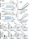
Som+ CeLC neurons are more excitable than PKCδ+ cells. A, Representative voltage traces of late-firing (top left) or regular-spiking (bottom left) PKCδ+ cells (black) or Som+ cells (blue) in response to depolarizing current injections. Right panel shows the number of spikes elicited as a function of the current injection amplitude; ****p < 0.0001, **p < 0.0036, two-way ANOVA. B–I, Latency to first spike (B, F), rheobase (C, G), Rin (D, H), and resting membrane potential (Vrest; E, I) for late-firing (B–E) and regular-spiking (F–I) neurons; **p = 0.0039, unpaired two-tailed t test; *p = 0.0308, Mann–Whitney U test; ***p = 0.0002, unpaired two-tailed t test with Welch’s correction. For PKCδ+ cells: n = 16–19 cells for late-firing and n = 35–36 regular-spiking. For Som+ cells: n = 9 for late-firing and n = 12 for regular-spiking. All values are expressed as mean ± SEM.
Figure 4.
Figure 4.
Voltage sag is indistinguishable in PKCδ+ and Som+ cells. Representative traces of late-firing (top) and regular-spiking (bottom) neurons in response to a 500-ms hyperpolarizing current injection, with PKCδ+ cells shown in black and Som+ cells in blue. Values are reported as mean ± SEM. For PKCδ+ cells: n = 18 cells for late-firing and n = 33 regular-spiking. For Som+ cells: n = 9 for late-firing and n = 13 for regular-spiking. Voltage sag = Vsag.
Figure 5.
Figure 5.
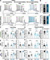
Accommodation is selective to PKCδ+, but not Som+, neurons. A, Representative voltage records of PKCδ+ accommodating (Ac, left) and non-accommodating (Na, center) cells and Som+ Na cells (right) for late-firing (top) regular-spiking (bottom) cells. Pink annotations depict ISI accommodation, green denotes spike amplitude accommodation, and purple shows AHP amplitude accommodation. B, The proportions of Na and Ac late-firing and regular-spiking PKCδ+ and Som+ cells. C–H, Spike amplitude accommodation (C, D), APD accommodation (E, F), and AHP amplitude accommodation (G, H) for late-firing (left) and regular-spiking (right) PKCδ+ and Som+ cells; ****p < 0.0001, ***p < 0.005, paired two-tailed t test; **p = 0.0034, unpaired two-tailed t test; *p = 0.0142, unpaired two-tailed t test with Welch’s correction. For PKCδ+ cells: n = 14–16 cells for late-firing and n = 33–36 regular-spiking. For Som+ cells: n = 9 for late-firing and n = 12–13 for regular-spiking. All values are expressed as mean ± SEM.
Figure 6.
Figure 6.
Slower repolarization in Som+, than in PKCδ+ neurons. A, Representative single action potentials (left) elicited by 5-ms depolarizing current injections, phase plots (right) and plots of the first derivatives as a function of time (middle) of late-firing (top) and regular-spiking (bottom) PKCδ+ (black) and Som+ (blue) neurons. Insets depict expanded timescales. Single action potential analyses for late-firing (B–G) and regular-spiking (H–M) PKCδ+ and Som+ neurons. Current (B, H) and voltage (C, I) thresholds to fire a single action potential. Action potential rise time (D, J), action potential decay time (E, K), APD (F, L), and AHP amplitudes (G, M); ****p < 0.0001, unpaired two-tailed t test with Welch’s correction; ####p < 0.0001, Mann–Whitney U test; *p = 0.0498, ***p = 0.0008, unpaired two-tailed t test. For PKCδ+ cells: n = 16 cells for late-firing and n = 31 regular-spiking. For Som+ cells: n = 9 for late-firing and n = 10 for regular-spiking. All values are expressed as mean ± SEM.
Figure 7.
Figure 7.
Spontaneous Som+ CeLC neurons are more active and fire longer action potentials than PKCδ+ cells. Representative spontaneous action potentials of PKCδ+ (A) and Som+ (B) neurons. Insets depict expanded timescales of single action potentials (B–G) and regular-spiking (H–L) PKCδ+ and Som+ neurons. Frequency of action potential firing (C), IS potential (D), capacitance (E), Rin (F), peak voltage (G), Vthreshold (H), rise (I), decay (J), duration (K), and AHP amplitudes (L) for PKCδ+ and Som+ cells; **p < 0.01, unpaired two-tailed t test with Welch’s correction; ***p < 0.001, unpaired two-tailed t test; ###p < 0.0002, unpaired two-tailed t test with Welch’s correction; ****p < 0.0001, unpaired two-tailed t test with Welch’s correction; ##p < 0.005, unpaired two-tailed t test. For PKCδ+ cells: n = 10–18 cells. For Som+ cells: n = 17–23 cells. All values are expressed as mean ± SEM.
Figure 8.
Figure 8.
Rostro-caudal distribution of PKCδ+ and Som+ neurons in the CeA. A, Representative low-magnification (left) and high-magnification (second to fifth panels) images of coronal CeA slices immunostained for PKCδ (PKCδ-IF, cyan), with cells positive for Som-tdTomato shown in red. Merged signals between PKCδ-IF and Som-tdTomato are shown in the fourth panels. Rightmost panels depict high-magnification images of areas delineated by the white box. Scale bars: 1 mm (left panels), 100 μm (middle panels), and 20 μm (right panels). B, Mean ± SEM number of cells positive for PKCδ (cyan), Som (red), or colabeled with both (white circles) in the capsular (CeC), lateral (CeL), or medial (CeM) subdivisions of the CeA, as well as the total number of positive cells, are shown as a function of the rostro-caudal distribution relative to bregma; n = 1–8 slices per rostro-caudal level from a total of 2–10 mice.
Figure 9.
Figure 9.
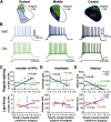
Firing responses in regular-spiking PKCδ+ neurons correlate with rostro-caudal anatomic location within the CeC. A, Schematics of rostral, middle, and caudal regions of the CeLC, with the CeL represented in green and purple and the CeC represented in blue and pink. B, Representative voltage traces of evoked firing responses in regular-spiking PKCδ+ neurons in the rostral, middle, and caudal CeC (top panel, blue) and CeL (bottom panel, green). C–E, Correlational plots between the number of evoked action potentials (C), rheobase (D), or latency to fire (E) and the rostro-caudal location of regular-spiking (top) and late-firing (bottom) cells. Prolonged (500 ms) depolarizing current injections of 140 and 240 pA were used to evoke repetitive firing in regular-spiking and late-firing cells, respectively. For regular-spiking neurons in the CeC, there was a positive correlation between the number of evoked action potentials and the rostro-caudal level (p = 0.0045, r2 = 0.5031, linear regression analysis) and a negative correlation between rheobase (p = 0.0036, r2 = 0.5193, linear regression analysis) and latency to first spike (p = 0.0118, r2 = 0.4858, linear regression analysis) with the rostro-caudal level. None of the measured parameters in the CeL and in late-firing cells in the CeC correlated with the rostro-caudal level. For CeL: n = 7 cells for late-firing and n = 18 for regular-spiking. For CeC: n = 10 for late-firing and n = 14 for regular-spiking.
Figure 10.
Figure 10.
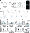
PKCδ+ and Som+ cells are morphologically distinct. A, Morphologic reconstruction of biocytin-filled cells in acute brain slices. CeLC cells were filled with biocytin during whole-cell patch-clamp recordings in acute amygdala slices. Representative images of biocytin-filled PKCδ+ and Som+ cells are shown in cyan in the right panel. Scale bars: 100 μm. B, Morphologic reconstruction of PKCδ+ (top) and Som+ biocytin-filled neurons. Scale bar: 100 μm. C, Sholl analysis for number of dendritic intersections as a function of radial distance from soma. D–F, Numbers (D), lengths (E), and spine densities (F) of primary (1°), secondary (2°), and tertiary (3°) dendrites for PKCδ+ and Som+ CeA cells; **p < 0.01, two-way ANOVA. G, I, Whole-cell membrane capacitance for late-firing (G) and regular-spiking (I) PKCδ+ and Som+ CeA cells. H, J, Correlational plots between the number of action potentials evoked in response to prolonged (500 ms) depolarizing current injections of either 140 pA (regular-spiking) or 240 pA (late-firing) and whole-cell membrane capacitance in late-firing (H) and regular-spiking (J) PKCδ+ (black) and Som+ (blue) CeA cells. A negative correlation was found in both late-firing (p = 0.0039, r2 = 0.29, linear regression analysis) and regular-spiking (p < 0.0001, r2 = 0.32, linear regression analysis) neurons. For PKCδ+ cells: n = 7 cells for morphology; n = 18–19 late-firing and n = 35–36 for regular-spiking. For Som+ cells: n = 6 cells for morphology; n = 9 late-firing and n = 13 for regular-spiking. All values are expressed as mean ± SEM.
Figure 11.
Figure 11.
Excitability differences between PKCδ+ and Som+ cells are occluded in the context of persistent pathologic pain. A, Cuff model of neuropathic pain used in electrophysiological experiments. Following placement of the sciatic nerve cuff, mice developed hypersensitivity to cold (acetone test), heat (Hargreaves test), and tactile (von Frey test) stimulation on hindpaws ipsilateral to nerve injury, compared with the contralateral hindpaws. Acute brain slices for electrophysiological experiments were collected following cuff placement in the sciatic nerve of Prkcd-Cre::Ai9 or Sst-Cre::Ai9 mice. B, Representative voltage recordings of late-firing (top) and regular-spiking (bottom) PKCδ+ (black) and Som+ (blue) cells in response to depolarizing current injections. Right panels show the number of spikes elicited as a function of the current injection amplitude. C, The number of action potentials elicited in response to 180- and 360-pA depolarizing current injections in late-firing (top) and regular-spiking (bottom) PKCδ+ (black) and Som+ (blue) CeA cells; ****p < 0.0001, ***p < 0.0002, *p = 0.0314, Mann–Whitney U test. n = 8–21 mice for behavioral tests. For PKCδ+ cells: n = 11–12 late-firing and n = 16–18 for regular-spiking. For Som+ cells: n = 7 late-firing and n = 20–21 for regular-spiking. All values are expressed as mean ± SEM.
Figure 12.
Figure 12.
Passive membrane and repetitive firing properties of PKCδ+ and Som+ cells following nerve injury. All data are reported as mean ± SEM. For PKCδ+ cells: n = 12 cells for late-firing and n = 18 regular-spiking. For Som+ cells: n = 6–7 for late-firing and n = 21 for regular-spiking. Vrest = resting membrane potential; Rin = input resistance.

References

    1. Aggleton JP (2000) The amygdala: a functional analysis. New York: Oxford University Press.
    1. Allen H, Bobnar H, Kolber BK (2020) Left and right hemispheric lateralization of the amygdala in pain. Prog Neurobiol. Advance online publication. Retrieved July 28, 2020. doi: 10.1016/j.pneurobio.2020.101891. - PMC - PubMed
    1. Amano T, Amir A, Goswami S, Paré D (2012) Morphology, PKCδ expression, and synaptic responsiveness of different types of rat central lateral amygdala neurons. J Neurophysiol 108:3196–3205. 10.1152/jn.00514.2012 - DOI - PMC - PubMed
    1. Avegno EM, Middleton JW, Gilpin NW (2019) Synaptic GABAergic transmission in the central amygdala (CeA) of rats depends on slice preparation and recording conditions. Physiol Rep 7:e14245. 10.14814/phy2.14245 - DOI - PMC - PubMed
    1. Bean BP (2007) The action potential in mammalian central neurons. Nat Rev Neurosci 8:451–465. 10.1038/nrn2148 - DOI - PubMed

LinkOut - more resources