. 2020 Nov 13;6(46):eabc5569.
doi: 10.1126/sciadv.abc5569. Print 2020 Nov.

Lack of nutritional immunity in diabetic skin infections promotes Staphylococcus aureus virulence

Affiliations

Lack of nutritional immunity in diabetic skin infections promotes Staphylococcus aureus virulence

Lance R Thurlow et al. Sci Adv. .

Abstract

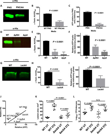
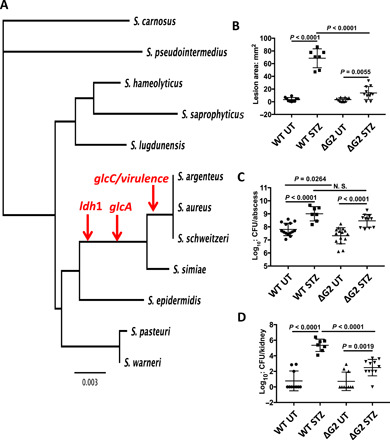
Elevated blood/tissue glucose is a hallmark feature of advanced diabetes, and people with diabetes are prone to more frequent and invasive infections with Staphylococcus aureus. Phagocytes must markedly increase glucose consumption during infection to generate and oxidative burst and kill invading bacteria. Similarly, glucose is essential for S. aureus survival in an infection and competition with the host, for this limited resource is reminiscent of nutritional immunity. Here, we show that infiltrating phagocytes do not express their high-efficiency glucose transporters in modeled diabetic infections, resulting in a diminished respiratory burst and increased glucose availability for S. aureus We show that excess glucose in these hyperglycemic abscesses significantly enhances S. aureus virulence potential, resulting in worse infection outcomes. Last, we show that two glucose transporters recently acquired by S. aureus are essential for excess virulence factor production and the concomitant increase in disease severity in hyperglycemic infections.


Figures

Fig. 1
Fig. 1. Diminished oxidative burst in hyperglycemic abscesses correlates with worse infection outcomes.
Untreated (UT) (N = 9 and N = 11) and STZ-treated (N = 11 and N = 8) mice were infected with 1 × 10⁷ CFU of the USA300 methicillin-resistant S. aureus (MRSA) strain LAC. WT-infected STZ-treated mice have a larger dermonecrotic lesion (A), increased abscess burden (B), and increased dissemination to the kidney (C) compared with similarly infected untreated mice. Pictures of dermonecrotic lesions from an untreated mouse (red arrows) (D) and a STZ-treated mouse (E) show enhanced dermonecrosis in hyperglycemic infection. Immunohistochemistry (IHC) was performed on tissues from untreated and STZ-treated infections using antibodies against iNOS, nitrotyrosine (Y-NO2), or GLUT-1 (red) and counterstained with 4′,6-diamidino-2-phenylindole (DAPI) (blue). (F) Tissues from both untreated and STZ-treated mice show iNOS staining. (G) However, while untreated mice exhibit robust Y-NO2 staining, this adduct is largely absent in tissue from STZ-treated animals. (H) Similarly, tissues from untreated mice show robust GLUT-1 staining that is mostly absent in tissue from STZ-treated animals. Photo credit: Lance Thurlow, University of Pittsburgh.
Fig. 2
Fig. 2. Agr is required for invasive infections in hyperglycemic mice and requires glucose for activation.
Glucose levels were measured in abscesses from untreated and STZ-treated animals and normalized to protein levels. (A) Abscesses from STZ-treated animals had significantly higher glucose levels than those from untreated animals. (B) S. aureus strain LAC harboring a plasmid with YPF linked to the RNAIII promoter (filled symbols) were grown in PNG (red symbols) or PNCAA (blue symbols). Fluorescence is only observed in strains grown in PNG (left axis) (B). (C) Relative RNAIII:YFP fluorescence normalized to OD660 at 10 hours shows greater induction of RNAIII in strains LAC and COL grown in PNG (n = 3). Normal (N = 9 and N = 11) and diabetic (N = 11 and N = 8) mice were infected with 1 × 10⁷ CFU of the USA300 MRSA strain LAC or LAC agr::Tn (n = 10 for untreated and N = 8 for STZ-treated). (D) Neither treated nor STZ-treated mice infected with agr::Tn formed necrotic lesions. (E) The agr::Tn abscesses had lower bacterial burdens than WT-infected mice in both untreated and STZ-treated mice; however, the reduced burden in STZ-treated mice was not as notable. (F) STZ-treated mice infected with agr::Tn had significantly less dissemination to the kidneys compared with STZ-treated mice infected with WT LAC.
Fig. 3
Fig. 3. Toxin production by S. aureus requires ATP generation from glycolysis.
(A) Representative Western blot showing Hla in spent media from LAC grown in PNG (left) or PNCAA (right). (B) Quantification of Hla normalized to OD660 in LAC grown in PNG or PNCAA (n = 3). AU, arbirtrary unit. (C)Quantification of intracellular ATP normalized to OD660 relative to LAC grown in PNG versus PNCAA (n = 3). (D) Representative Hla Western blots from culture supernatants from LAC (left), LAC ΔpfkA (center), and LAC Δpyk (right) grown in PNMix. (E) Quantification of Western blots from LAC, LAC ΔpfkA, and LAC Δpyk (n = 3). (F) Quantification of intracellular ATP normalized to OD660 relative to LAC. (G) Representative Hla Western blots of culture supernatant from LAC (left) and LAC ΔackA (right) grown in PNG. (H) Quantification of α-toxin Western blots from LAC and LAC ΔackA (n = 3). (I) Quantification of intracellular ATP normalized to OD660 relative to LAC (n = 3). (J) Regression analysis revealed a significant correlation between normalized intracellular ATP and the amount of normalized excreted Hla (R2 = 0.7, nonzero slope, P = 0.019). Untreated and STZ-treated mice were infected with 107 CFU of WT LAC or LAC ΔackA (WT UT, n = 10; WT STZ, n = 7; ΔackA UT, n = 10; and ΔackA STZ, n = 11). (K) Untreated mice infected with WT or ΔackA have similar lesion sizes. STZ-treated mice infected with ΔakcA have significantly larger lesion sizes than similarly infected untreated mice, but smaller lesion sizes than STZ-treated mice infected with WT (K). (L) STZ-treated mice infected with WT have significantly increased bacterial burden in the subcutaneous abscess compared with similarly infected untreated mice. STZ-treated mice infected with ΔackA have significantly increased bacterial burden in the subcutaneous abscess compared with similarly infected untreated mice (L). There is no significant difference in abscess burden in STZ-treated mice infected with WT or ΔackA (L). N.S., not significant.
Fig. 4
Fig. 4. Reducing blood glucose in hyperglycemic mice with phlorizin reverses infection severity.
Mice were treated with phlorizin (Phlo) 1 day before infection and throughout the course of infection to lower blood glucose levels without influencing insulin signaling. (A) STZ-treated mice treated with phlorizin had smaller lesions than mice treated with STZ alone, but larger lesions than untreated mice. (B) Infected STZ-treated mice display significant weight loss that is reversed with phlorizin. (C) STZ-treated mice that were further treated with phlorizin display lower abscess burdens than mice treated with STZ-alone, but higher burdens than untreated mice. Phlorizin treatment resulted in decreased dissemination to the peripheral organs in STZ-treated mice including the kidneys (D), liver (E), and heart (F). (G) Quantitative real-time PCR S. aureus isolated from untreated, STZ-treated, and dual STZ phlorizin abscesses showed significantly increased expression (P < 0.05) of hla, psmα, and aur in STZ-treated mice that is reversed by phlorizin.
Fig. 5
Fig. 5. Glucose transporters are essential for ATP and toxin production.
(A) Representative Hla Western blot from supernatants of WT LAC, LAC ΔG4 (lacking all glucose transporters), and LAC ΔG2 (lacking the recently acquired GlcC and GlcA transporters) grown in PNG. (B) Quantification of Hla Western blots (n = 3) showing significantly decreased Hla production by LAC ΔG2 and LAC ΔG4 compared to WT LAC. (C) The LAC ΔG4 (lacking all glucose transporters) and LAC ΔG2 have reduced intracellular ATP compared to WT LAC when grown in PNG. (D) Quantitative real-time PCR from WT LAC and LAC ΔG4 grown in PNG shows significant decreases in aur, psmα, and hla transcripts in LAC ΔG4 (n = 3). (E) Quantitative real-time PCR from WT LAC and LAC ΔG4 isolated from STZ-treated mice showed reduced transcript levels of hla, psmα, and aur in LAC ΔG4. (F) Day 7 lesion sizes measured from untreated mice infected with LAC ΔG4 were virtually undetectable, and abscesses in STZ-treated mice infected with LAC ΔG4 also rarely had small lesions. (G) LAC ΔG4 is attenuated in untreated and STZ-treated mice compared with WT LAC but has higher CFU in STZ-treated animals.
Fig. 6
Fig. 6. The evolutionary acquisition of glucose transporters by S. aureus coincides with acquisition of virulence factors.
A phylogenetic tree based on 16S ribosomal DNA sequences showing the relation of S. aureus to other staphylococcal species. Red arrows show where S. aureus and other related species evolutionarily acquired lactate dehydrogenase 1 (ldh1), glucose transporter A (glcA), glucose transporter C (glcC), and virulence factors including α-, β-, and γ-hemolysins (A). Untreated and STZ-treated mice were infected with 1 × 10⁷ CFU of WT and ∆G2 (WT UT, n = 10; WT STZ, n = 7 to 8; ∆G2 UT, n = 10; and ∆G2 STZ, n = 11). STZ-treated mice infected with WT had larger lesion sizes than infected untreated mice. STZ-treated mice infected with ΔG2 had smaller lesion sizes than WT infected STZ-treated mice (B). (C) STZ-treated mice infected with WT or ΔG2 had significantly higher abscess bacterial burdens than their untreated infected controls. (D) STZ-treated mice infected with WT displayed increased dissemination to the kidneys compared to similarly infected untreated mice. STZ-treated mice infected with ΔG2 had increased dissemination to the kidneys compared to similarly infected untreated mice (D).

References

    1. Abu-Ashour W., Twells L., Valcour J., Randell A., Donnan J., Howse P., Gamble J.-M., The association between diabetes mellitus and incident infections: A systematic review and meta-analysis of observational studies. BMJ Open Diabetes Res. Care 5, e000336 (2017). - PMC - PubMed
    1. Abu-Ashour W., Twells L. K., Valcour J. E., Gamble J.-M., Diabetes and the occurrence of infection in primary care: A matched cohort study. BMC Infect. Dis. 18, 67 (2018). - PMC - PubMed
    1. Lipsky B. A., Tabak Y. P., Johannes R. S., Vo L., Hyde L., Weigelt J. A., Skin and soft tissue infections in hospitalised patients with diabetes: Culture isolates and risk factors associated with mortality, length of stay and cost. Diabetologia 53, 914–923 (2010). - PubMed
    1. Smit J., Søgaard M., Schønheyder H. C., Nielsen H., Frøslev T., Thomsen R. W., Diabetes and risk of community-acquired Staphylococcus aureus bacteremia: A population-based case-control study. Eur. J. Endocrinol. 174, 631–639 (2016). - PubMed
    1. Jneid J., Cassir N., Schuldiner S., Jourdan N., Sotto A., Lavigne J. P., la Scola B., Exploring the microbiota of diabetic foot infections with culturomics. Front. Cell. Inf. Microbiol. 8, 282 (2018). - PMC - PubMed

Publication types