. 2020 Nov;8(2):e001367.
doi: 10.1136/jitc-2020-001367.

Combination of gemcitabine and anti-PD-1 antibody enhances the anticancer effect of M1 macrophages and the Th1 response in a murine model of pancreatic cancer liver metastasis

Affiliations

Combination of gemcitabine and anti-PD-1 antibody enhances the anticancer effect of M1 macrophages and the Th1 response in a murine model of pancreatic cancer liver metastasis

Tuyen Thuy Bich Ho et al. J Immunother Cancer. 2020 Nov.

Abstract

Background: Pancreatic ductular adenocarcinoma (PDAC) is among the most dreadful of malignancies, in part due to the lack of efficacious chemotherapy. Immune checkpoint inhibitors, including anti-programmed cell death 1 (anti-PD-1) antibodies, are novel promising forms of systemic immunotherapy. In the current study, we assessed whether gemcitabine (GEM) combined with anti-PD-1 antibody treatment was efficacious as immunochemotherapy for advanced PDAC using a murine model of liver metastasis.

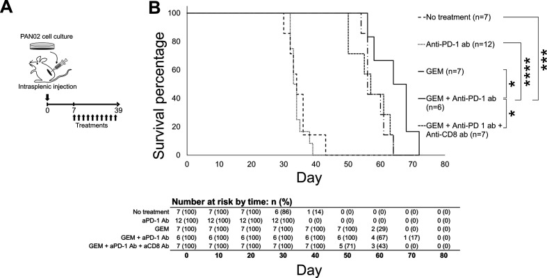
Methods: The murine model of PDAC liver metastasis was established by intrasplenically injecting the murine pancreatic cancer cell line PAN02 into immunocompetent C57BL/6J mice. The mice were treated with an anti-PD-1 antibody, GEM, or a combination of GEM plus anti-PD-1 antibody, and compared with no treatment (control); liver metastases, immune cell infiltration, gene expression, immune cell response phenotypes, and overall survival were investigated.

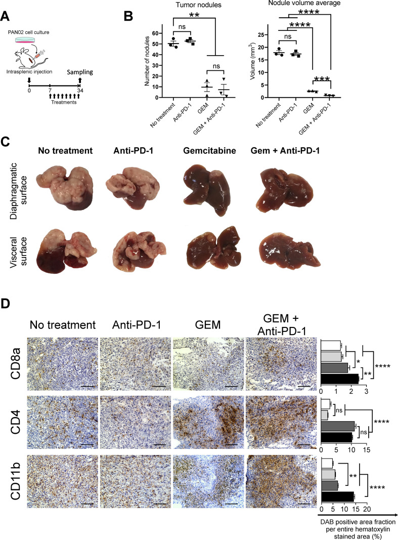
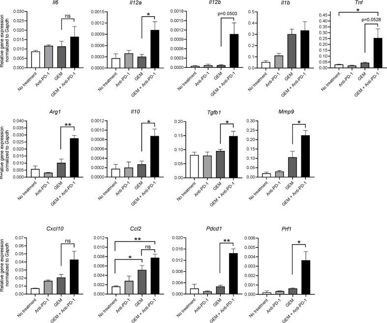
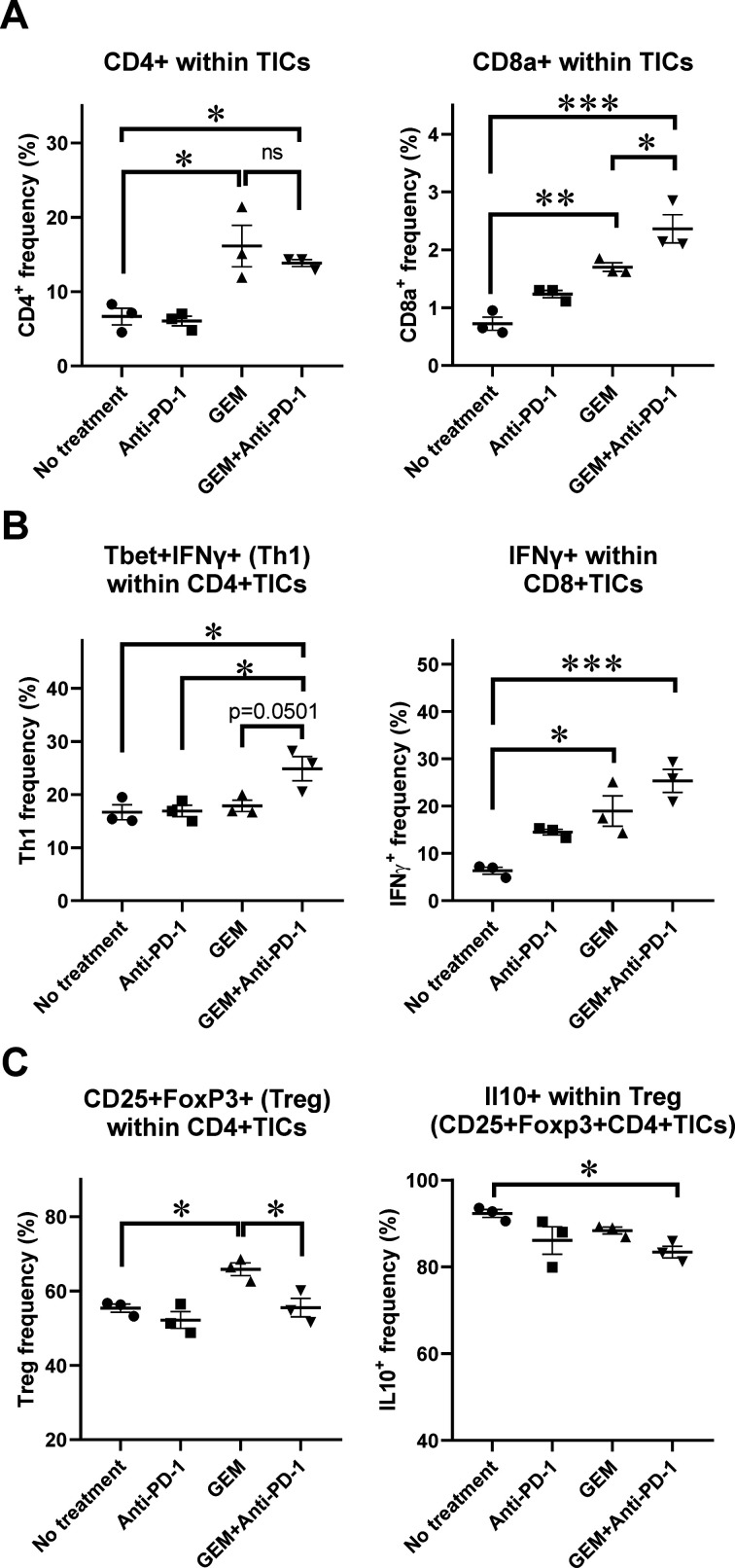
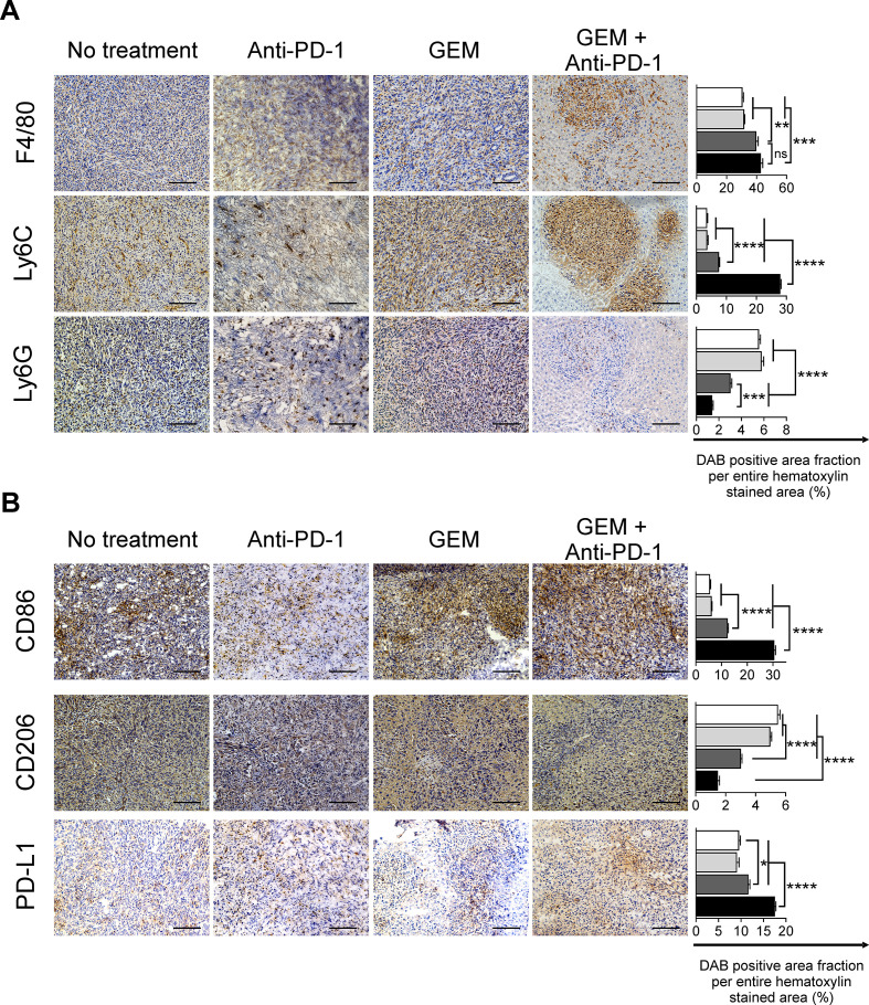
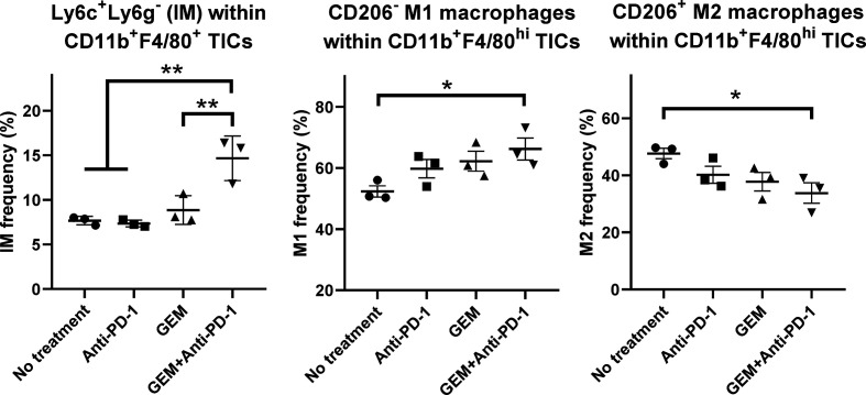
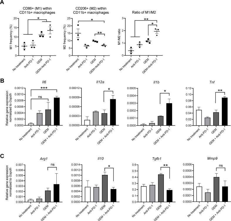
Results: In the metastatic tumor tissues of mice treated with GEM plus anti-PD-1 antibody, we observed the increased infiltration of Th1 lymphocytes and M1 macrophages. Gene expression profile analysis of peripheral blood cells obtained from mice treated with GEM plus anti-PD-1 antibody clearly highlighted T cell and innate immune signaling pathways. Survival of PDAC liver metastasis mice was significantly prolonged by the combination therapy (median survival, 66 days) when compared with that of GEM alone treatment (median survival, 56 days). Expanded lymphocytes, which were isolated from the splenocytes of PDAC liver metastasis mice treated with GEM plus anti-PD-1 antibody, had an increased number of M1 macrophages.

Conclusion: The combination of anti-PD-1 antibody immunotherapy with GEM was beneficial to treat a murine model of PDAC liver metastasis by enhancing the immune response mediated by Th1 lymphocytes and M1 macrophages and was associated with CD8+ T cells.

Keywords: combination; drug therapy; immunity; immunotherapy; macrophages; tumor microenvironment.


Conflict of interest statement

Competing interests: None declared.

Figures

Figure 1
Figure 1
The response in pancreatic ductular adenocarcinoma (PDAC) liver metastasis tumors of mice treated with gemcitabine (GEM) and anti-programmed cell death 1 (anti-PD-1) Ab. (A) Experimental schedule showing the treatments for PDAC liver metastasis mice: no treatment (phosphate-buffered saline), anti-PD-1 Ab, GEM, and GEM plus anti-PD-1 Ab. Treatment was administered twice a week from day 7 to day 33. Tumor tissues were obtained and stained immunohistochemically on day 34 (n=3 per group). (B) Number of liver metastasis tumor nodules and averaged nodule volumes and (C) macroscopic images of tumors are shown; (B) bars represent mean±SEM; Student’s t-test was performed as statistical analysis. (D) Immunohistochemical analysis of tumors for CD8a+, CD4+, and CD11b+ inflammatory cells. Magnification: ×200; bars: 100 µm. Quantification of cell infiltration by using ImageJ (each area analyzed=1.576 mm2, three different areas were analyzed for each sample). White bar: no treatment; light gray bar: anti-PD-1 Ab; dark gray bar: GEM; black bar: GEM plus anti-PD-1 Ab; bars represent mean±SEM; one-way analysis of variance followed by Tukey’s HSD post hoc test was performed as statistical analysis; *p<0.05, **p<0.01, ***p<0.001, ****p<0.0001. DAB, 3,3'-diaminobenzidine; HSD, honestly significant difference.
Figure 2
Figure 2
Gene expression analysis of tumor-infiltrating inflammatory cells (TICs) from pancreatic ductular adenocarcinoma (PDAC) liver metastasis model mice by qRT-PCR. PDAC liver metastasis mice received a single dose of the indicated treatment on day 28 and tumor tissues were obtained 2 days later for TIC isolation, followed by RNA extraction. Quantitative RT-PCR showing the expression of M1 macrophage-related cytokine genes (Il6, Il12a, Il12b, Il1b, and Tnf), M2 macrophage-related genes (Arg1, Il10, Tgfb1, and Mmp9), proinflammatory chemokines (Cxcl10 and Ccl2), and T-cell activation markers (Pdcd1 and Prf1); n=3; bars represent mean±SEM; Student’s t-test was used for statistical analysis; *p<0.05, **p<0.01. GEM, gemcitabine; PD-1, programmed cell death 1; qRT-PCR, quantitative real-time polymerase chain reaction.
Figure 3
Figure 3
Flow cytometry (FCM) analysis of tumor-infiltrating inflammatory cells (TICs) for lymphoid-lineage cells. Pancreatic ductular adenocarcinoma liver metastasis mice received a single dose of the indicated treatment on day 28 and tumor tissues were obtained 2 days later for TIC isolation followed by FCM analysis; n=3. (A) CD4+ and CD8a+ cells within TICs. (B) Th1 CD4+ cells expressing T-bet and interferon (IFN)-γ; IFN-γ-secreting cells within CD8+ TICs. (C) Regulatory T cell phenotype, CD4+CD25+FoxP3+ cells, and interleukin (IL)-10+-producing cells within Treg cells. (A–C) Bars represent mean±SEM; one-way analysis of variance followed by Tukey’s HSD post hoc test was performed as statistical analysis; *p<0.05, **p<0.01, ***p<0.001, ****p<0.0001. GEM, gemcitabine. PD-1, programmed cell death 1.
Figure 4
Figure 4
Immunohistochemical analysis of pancreatic ductular adenocarcinoma liver metastasis tumors for myeloid-lineage cells. Treatments were conducted twice a week from day 7 to day 33; tumors were obtained on day 34 and immunohistochemically analyzed for (A) F4/80+, Ly6C+, and Ly6G+, and (B) CD86+, CD206+ infiltrating cells, and programmed cell death-ligand 1 (PD-L1)+ for the indicated treatment. Magnification: ×200; bars: 100 µm. (A, B) Quantification of cell infiltration by using ImageJ (each area analyzed=1.576 mm2, 3 different areas were analyzed for each sample). White bar: no treatment; light gray bar: anti-PD-1 Ab; dark gray bar: gemcitabine (GEM); black bar: GEM plus anti-PD-1 Ab. Bars represent mean±SEM; one-way analysis of variance followed by Tukey’s HSD post hoc test was performed as statistical analysis; *p<0.05, **p<0.01, ***p<0.001, ****p<0.0001.
Figure 5
Figure 5
Flow cytometry (FCM) analysis of tumor-infiltrating inflammatory cells (TICs) for myeloid-lineage cells. Pancreatic ductular adenocarcinoma liver metastasis mice received the following treatments: no treatment, anti-programmed cell death 1 (anti-PD-1) Ab, gemcitabine (GEM), or GEM plus anti-PD-1 Ab on day 28 and tumor tissues were obtained 2 days later for TIC isolation and FCM; n=3, bars represent mean±SEM. The following cell populations were analyzed: Ly6C+Ly6G− inflammatory monocytes within CD11b+F4/80+ TICs, CD206− M1 macrophages within CD11b+F4/80high TICs, and CD206+ M2 macrophages within CD11b+F4/80high TICs. One-way analysis of variance followed by Tukey’s HSD post hoc test was performed as statistical analysis; *p<0.05, **p<0.01, ***p<0.001, ****p<0.0001.
Figure 6
Figure 6
Survival of pancreatic ductular adenocarcinoma (PDAC) liver metastasis model mice that underwent treatment. (A) Experimental schedule showing treatment timing: the PDAC liver metastasis mice received treatment 10 times, twice a week from day 7 to day 39, and survival percentage was monitored until day 72. (B) Survival curves of PDAC liver metastasis mice that received: no treatment (only phosphate-buffered saline; n=7), anti-programmed cell death 1 (anti-PD-1) Ab (n=12), gemcitabine (GEM) (n=7), GEM plus anti-PD-1 Ab (n=6), or GEM plus anti-PD-1 Ab plus anti-CD8 Ab (n=7). The log-rank test was performed to obtain the p values: no treatment/anti-PD-1 Ab=0.492723; no treatment/GEM=0.000172; no treatment/GEM plus anti-PD-1 Ab=0.000446; anti-PD-1 Ab/GEM=0.000034; anti-PD-1 Ab/GEM plus anti-PD-1 Ab=0.000098; GEM/GEM plus anti-PD-1 Ab=0.034672; and GEM plus anti-PD-1 Ab/GEM plus anti-PD-1 Ab plus anti-CD8 Ab=0.0345.
Figure 7
Figure 7
In vitro macrophage polarization assay. Pancreatic ductular adenocarcinoma liver metastasis mice received a single dose of the indicated treatment on day 28 and splenocytes were obtained on day 30. The splenocytes were activated by coculture with irradiated PAN02 cells; after 7 days, activated splenocytes were harvested and cocultured with bone marrow-derived macrophages. After 48 hours, macrophages were collected for (A) flow cytometry analysis of CD11b, CD86, and CD206; and total RNA was isolated for qRT-PCR analysis of (B) Il6, Il12a, Il1b, and Tnf (M1 macrophage-related genes) or (C) Arg1, Il10, Tgfb1, and Mmp9 (M2 macrophage-related genes). (A–C) n=3; bars represent mean±SEM; Student’s t-test was performed for statistical analysis; *p<0.05, **p<0.01, ***p<0.001. GEM, gemcitabine; PD-1, programmed cell death 1.

References

    1. Ryan DP, Hong TS, Bardeesy N. Pancreatic adenocarcinoma. N Engl J Med 2014;371:1039–49. 10.1056/NEJMra1404198 - DOI - PubMed
    1. Iacobuzio-Donahue CA, Fu B, Yachida S, et al. . Dpc4 gene status of the primary carcinoma correlates with patterns of failure in patients with pancreatic cancer. J Clin Oncol 2009;27:1806–13. 10.1200/JCO.2008.17.7188 - DOI - PMC - PubMed
    1. Komura T, Sakai Y, Harada K, et al. . Inflammatory features of pancreatic cancer highlighted by monocytes/macrophages and CD4+ T cells with clinical impact. Cancer Sci 2015;106:672–86. 10.1111/cas.12663 - DOI - PMC - PubMed
    1. Clark CE, Hingorani SR, Mick R, et al. . Dynamics of the immune reaction to pancreatic cancer from inception to invasion. Cancer Res 2007;67:9518–27. 10.1158/0008-5472.CAN-07-0175 - DOI - PubMed
    1. Sahin IH, Askan G, Hu ZI, et al. . Immunotherapy in pancreatic ductal adenocarcinoma: an emerging entity? Ann Oncol 2017;28:2950–61. 10.1093/annonc/mdx503 - DOI - PMC - PubMed

Publication types

MeSH terms