. 2020 Nov 13;11(1):5768.
doi: 10.1038/s41467-020-18910-x.

Complex multicomponent patterns rendered on a 3D DNA-barrel pegboard

Affiliations

Complex multicomponent patterns rendered on a 3D DNA-barrel pegboard

Shelley F J Wickham et al. Nat Commun. .

Abstract

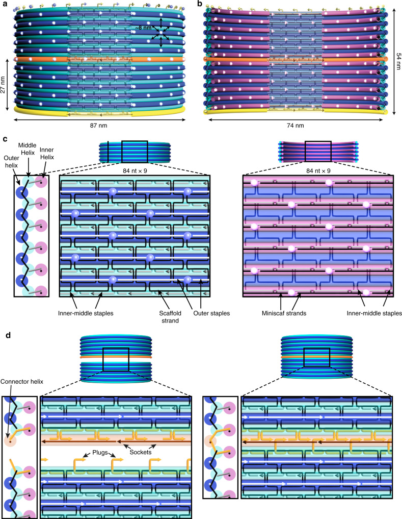
DNA origami, in which a long scaffold strand is assembled with a many short staple strands into parallel arrays of double helices, has proven a powerful method for custom nanofabrication. However, currently the design and optimization of custom 3D DNA-origami shapes is a barrier to rapid application to new areas. Here we introduce a modular barrel architecture, and demonstrate hierarchical assembly of a 100 megadalton DNA-origami barrel of ~90 nm diameter and ~250 nm height, that provides a rhombic-lattice canvas of a thousand pixels each, with pitch of ~8 nm, on its inner and outer surfaces. Complex patterns rendered on these surfaces were resolved using up to twelve rounds of Exchange-PAINT super-resolution microscopy. We envision these structures as versatile nanoscale pegboards for applications requiring complex 3D arrangements of matter, which will serve to promote rapid uptake of this technology in diverse fields beyond specialist groups working in DNA nanotechnology.


Conflict of interest statement

The authors declare no competing interests.

Figures

Fig. 1
Fig. 1. Schematic of coaxially stacked dimer of DNA-origami barrels with ~90-nm outer diameter.
Barrels consist of three layers of circular double helices arranged with honeycomb-lattice spacing: a measured from helix mid-point, outer helices (dark blue) are 84 nm in diameter, inner-middle helices (cyan) are 81.5 nm in diameter, b inner helices (magenta) are 76.5 nm in diameter. Including helix thickness of 2.6 nm gives external barrel diameter of 87 nm and height of 27 nm, with internal cavity of 74 nm. c Zoom-in of barrel wall cross-section (left panel), outer surface (middle panel), and inner surface (right panel) for central cut-away region. On the outer surface (left), scaffold routing is rendered as black pipes, and only runs through the outer and middle helix layers. Outer staple strands are rendered as white pipes, and blue fluorescent spheres indicate 3′ ends of outer staples, which form the rhombic-lattice distribution of outside pixel sites. Inner-middle staples are rendered as gray pipes on both surfaces. On the inner surface (right), there is no M13 scaffold DNA. Inner staple strands hybridize to short miniscaf strands, which are rendered as black pipes (left panel). Rhombic-lattice distribution of inside pixel sites, which are 3′ ends of inner miniscaf strands, is rendered as magenta fluorescent spheres. d Zoom-in detail of the staple strands that mediate coaxial stacking between monomers, rendered as orange pipes. Staple-strand extensions (plugs) on the top of the interface hybridize to connector strands (dark orange), leaving single-stranded regions (sockets), which plugs from the second barrel hybridize to. In zoom-out view a, b two orthogonal sets of connector strands are rendered in yellow and orange.
Fig. 2
Fig. 2. DNA-origami barrel monomers.
a Agarose gel electrophoresis of five barrel designs. Lanes 4–8 are barrels with diameter–height in nm: 30–27, 30–65, 60–30, 90–19, 90–23. Controls are 1-kb ladder (250–10,000 bp, lanes 1, 11; reference bands marked for 1, 3, 6-kb dsDNA), and unfolded scaffold (7308-nt lanes 2, 10; 8634-nt lanes 3, 9). b Design and molecular microscopy of barrel monomers excised from agarose gels as in a and recovered by centrifugation. Imaging was performed via (middle) DNA-PAINT super-resolution fluorescence microscopy or (right) uranyl-formate negative-stain TEM. To prepare for DNA-PAINT imaging, monomers first were functionalized with 6, 12, 18 biotins on their bottom inner helices for upright attachment to streptavidin-coated glass surfaces, and 6, 12, 18 docking handles on one of the outer helices (for 30, 60, 90-nm diameter barrels, respectively; indicated by red dots in design schematic). DNA-PAINT images are given for a single structure (left) and as a summed image (right) of N = 726, N = 16, N = 27 particles for the 30, 60, 90-nm barrels, respectively. Design and measured diameters of barrels are indicated on panels for both imaging methods. Scale bars 50 nm.
Fig. 3
Fig. 3. DNA-origami barrel polymers assembled from repeating α and β monomers.
a Models of barrel polymers illustrating placement of DNA-PAINT docking handles. 30-nm barrel design: α monomer with two DNA-PAINT rings spaced 50 nm apart, and αβ repeating height of 125 nm; 60-nm barrel design: α monomer with one DNA-PAINT ring and αβ repeating height of 58 nm; 90-nm barrel design: α monomer with one DNA-PAINT ring and αβ repeating height of 42 nm. b DNA-PAINT images of representative polymers with corresponding models listing designed spacings above. (Scale bars in a and b are 50 nm). c Histograms of measured spacings of DNA-PAINT rings. d Model of 90-nm barrel αβα trimer with four DNA-PAINT rings for each monomer, showing DNA-PAINT images of individual particles (colors for α: cyan, blue, magenta, yellow, colors for β: yellow, green, red, cyan). e Composite sum image (outer ring docking sites: X4, X5, X6, X1, X2, X3, X4, X5, X6, Supplementary Table 9). 3D model (left) shows designed ring spacing of 17 and 26 nm. f Histogram of distances between DNA-PAINT rings, measured ring spacing of 19 ± 10 and 27 ± 10 m. g Negative-stain TEM images of lipid nanotubes reconstituted within the interior of 90-nm barrel polymers. Scale bars are 50 nm.
Fig. 4
Fig. 4. Molecular microscopy of DNA-origami barrel decamers patterned with complex features (Supplementary Movies 1–6).
a Model of decamer, showing all possible pixel locations for DNA-PAINT docking handles. b Model of designed pattern of docking handles, for flattened and side-view (orthographic) of decamer. The design includes both outer and inner pixels. Pixels are colored by the 12 rounds of Exchange-PAINT (left) or grouped as four pseudocolors for ease of visualization (right). Model pixel size represents DNA-PAINT FWHM-resolution. c TEM of sideways decamer. Inset, TEM of upright monomer. d Model of designed placement of DNA-PAINT docking handles by pseudocolour, perspective view. e Exchange-PAINT image of a field of upright patterned particles alongside a few registration particles, with volume rendering. f Composite sum image with surface rendering. Inset, view from above. g Computationally arranged panel of selected particles with surface rendering. h Zoomed-in view seven particles boxed in g with surface rendering. i, j, Zoomed-in volume rendered particles boxed in e. Scale bars are 50 nm for c–f and are 500 nm for g, h.

References

    1. Seeman NC. Nanomaterials Based on DNA. Annu. Rev. Biochem. 2010;79:65–87. doi: 10.1146/annurev-biochem-060308-102244. - DOI - PMC - PubMed
    1. Wagenbauer KF, Sigl C, Dietz H. Gigadalton-scale shape-programmable DNA assemblies. Nature. 2017;552:78–83. doi: 10.1038/nature24651. - DOI - PubMed
    1. Tikhomirov G, Petersen P, Qian L. Fractal assembly of micrometre-scale DNA origami arrays with arbitrary patterns. Nature. 2017;552:67–71. doi: 10.1038/nature24655. - DOI - PubMed
    1. Wei B, Dai M, Yin P. Complex shapes self-assembled from single-stranded DNA tiles. Nature. 2012;485:623–626. doi: 10.1038/nature11075. - DOI - PMC - PubMed
    1. Ong LL, et al. Programmable self-assembly of three-dimensional nanostructures from 10,000 unique components. Nature. 2017;552:72–77. doi: 10.1038/nature24648. - DOI - PMC - PubMed

Publication types