

APOE-ε4-related differences in left thalamic microstructure in cognitively healthy adults
- PMID: 33188215
- PMCID: PMC7666117
- DOI: 10.1038/s41598-020-75992-9
APOE-ε4-related differences in left thalamic microstructure in cognitively healthy adults
Abstract
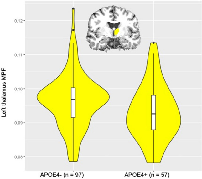
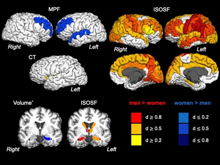
APOE-ε4 is a main genetic risk factor for developing late onset Alzheimer's disease (LOAD) and is thought to interact adversely with other risk factors on the brain. However, evidence regarding the impact of APOE-ε4 on grey matter structure in asymptomatic individuals remains mixed. Much attention has been devoted to characterising APOE-ε4-related changes in the hippocampus, but LOAD pathology is known to spread through the whole of the Papez circuit including the limbic thalamus. Here, we tested the impact of APOE-ε4 and two other risk factors, a family history of dementia and obesity, on grey matter macro- and microstructure across the whole brain in 165 asymptomatic individuals (38-71 years). Microstructural properties of apparent neurite density and dispersion, free water, myelin and cell metabolism were assessed with Neurite Orientation Density and Dispersion (NODDI) and quantitative magnetization transfer (qMT) imaging. APOE-ε4 carriers relative to non-carriers had a lower macromolecular proton fraction (MPF) in the left thalamus. No risk effects were present for cortical thickness, subcortical volume, or NODDI indices. Reduced thalamic MPF may reflect inflammation-related tissue swelling and/or myelin loss in APOE-ε4. Future prospective studies should investigate the sensitivity and specificity of qMT-based MPF as a non-invasive biomarker for LOAD risk.
Conflict of interest statement
The authors declare no competing interests.
Figures

Similar articles
-
Interactive effect of age and APOE-ε4 allele load on white matter myelin content in cognitively normal middle-aged subjects.Neuroimage Clin. 2019;24:101983. doi: 10.1016/j.nicl.2019.101983. Epub 2019 Aug 16. Neuroimage Clin. 2019. PMID: 31520917 Free PMC article.
-
Modulation on brain gray matter activity and white matter integrity by APOE ε4 risk gene in cognitively intact elderly: A multimodal neuroimaging study.Behav Brain Res. 2017 Mar 30;322(Pt A):100-109. doi: 10.1016/j.bbr.2017.01.027. Epub 2017 Jan 17. Behav Brain Res. 2017. PMID: 28108320
-
Genetic risk of dementia modifies obesity effects on white matter myelin in cognitively healthy adults.Neurobiol Aging. 2020 Oct;94:298-310. doi: 10.1016/j.neurobiolaging.2020.06.014. Epub 2020 Jun 29. Neurobiol Aging. 2020. PMID: 32736120
-
White matter microstructure is altered in cognitively normal middle-aged APOE-ε4 homozygotes.Alzheimers Res Ther. 2018 May 24;10(1):48. doi: 10.1186/s13195-018-0375-x. Alzheimers Res Ther. 2018. PMID: 29793545 Free PMC article.
-
Apolipoprotein E ε4 genotype status is not associated with neuroimaging outcomes in a large cohort of HIV+ individuals.J Neurovirol. 2016 Oct;22(5):607-614. doi: 10.1007/s13365-016-0434-7. Epub 2016 Mar 28. J Neurovirol. 2016. PMID: 27021072 Free PMC article.
Cited by
-
Quantitative magnetic resonance imaging in Alzheimer's disease: a narrative review.Quant Imaging Med Surg. 2025 Apr 1;15(4):3641-3664. doi: 10.21037/qims-24-1602. Epub 2025 Mar 28. Quant Imaging Med Surg. 2025. PMID: 40235823 Free PMC article. Review.
-
Effects of APOE2 and APOE4 on brain microstructure in older adults: modification by age, sex, and cognitive status.Alzheimers Res Ther. 2024 Jan 11;16(1):7. doi: 10.1186/s13195-023-01380-w. Alzheimers Res Ther. 2024. PMID: 38212861 Free PMC article.
-
Effects of obesogenic diet and 17β-estradiol in female mice with APOE 3/3, 3/4, and 4/4 genotypes.Front Aging Neurosci. 2024 Sep 13;16:1415072. doi: 10.3389/fnagi.2024.1415072. eCollection 2024. Front Aging Neurosci. 2024. PMID: 39347015 Free PMC article.
-
Longitudinal effects of sex differences and apolipoprotein E genotype on white matter engagement among elderly.Brain Commun. 2025 Jul 17;7(4):fcaf278. doi: 10.1093/braincomms/fcaf278. eCollection 2025. Brain Commun. 2025. PMID: 40740432 Free PMC article.
-
Effects of non-modifiable risk factors of Alzheimer's disease on intracortical myelin content.Alzheimers Res Ther. 2022 Dec 31;14(1):202. doi: 10.1186/s13195-022-01152-y. Alzheimers Res Ther. 2022. PMID: 36587227 Free PMC article.
References
-
- World Health Organisation. Dementia Factsheet. (https://www.who.int/news-room/fact-sheets/detail/dementia, 2019).
Publication types
MeSH terms
Substances
Grants and funding
LinkOut - more resources
Full Text Sources
Miscellaneous
