

Independent Contributions of Dorsolateral Prefrontal Structure and Function to Working Memory in Healthy Older Adults
- PMID: 33188384
- PMCID: PMC7869098
- DOI: 10.1093/cercor/bhaa322
Independent Contributions of Dorsolateral Prefrontal Structure and Function to Working Memory in Healthy Older Adults
Abstract
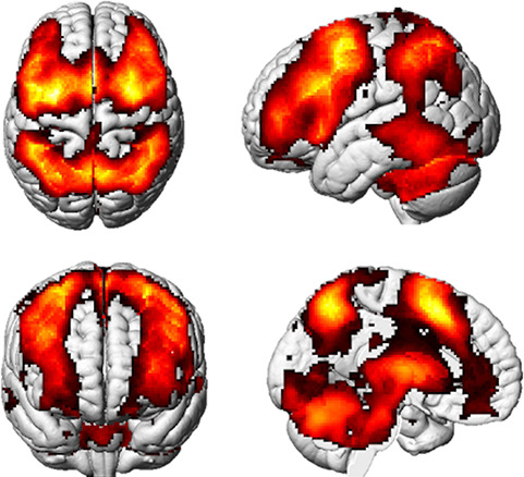
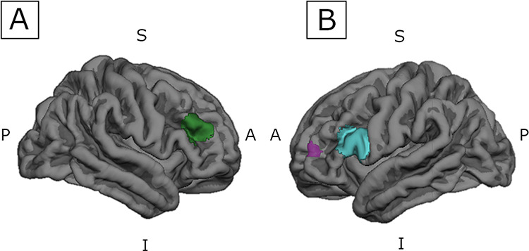
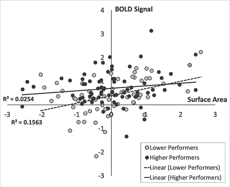
Age-related differences in dorsolateral prefrontal cortex (DLPFC) structure and function have each been linked to working memory. However, few studies have integrated multimodal imaging to simultaneously investigate relationships among structure, function, and cognition. We aimed to clarify how specifically DLPFC structure and function contribute to working memory in healthy older adults. In total, 138 participants aged 65-88 underwent 3 T neuroimaging and were divided into higher and lower groups based on a median split of in-scanner n-back task performance. Three a priori spherical DLPFC regions of interest (ROIs) were used to quantify blood-oxygen-level-dependent (BOLD) signal and FreeSurfer-derived surface area, cortical thickness, and white matter volume. Binary logistic regressions adjusting for age, sex, education, and scanner type revealed that greater left and right DLPFC BOLD signal predicted the probability of higher performing group membership (P values<.05). Binary logistic regressions also adjusting for total intracranial volume revealed left DLPFC surface area that significantly predicted the probability of being in the higher performing group (P = 0.017). The left DLPFC BOLD signal and surface area were not significantly associated and did not significantly interact to predict group membership (P values>.05). Importantly, this suggests BOLD signal and surface area may independently contribute to working memory performance in healthy older adults.
Keywords: cognitive aging; dorsolateral prefrontal cortex; multimodal neuroimaging; structural and functional magnetic resonance imaging; working memory.
© The Author(s) 2020. Published by Oxford University Press. All rights reserved. For permissions, please e-mail: journals.permission@oup.com.
Figures
References
-
- Aiken LS, West SG, Reno RR. 1991. Multiple regression: testing and interpreting interactions. Nachdr. ed. Newbury Park, California: SAGE.
-
- Bachelard HS 1979. Brain energy metabolism. Biochem Soc Trans. 7:264–264.
-
- Baddeley A 1992. Working memory. Science. 255:556–559. - PubMed
-
- Barnes J, Ridgway GR, Bartlett J, Henley SMD, Lehmann M, Hobbs N, Clarkson MJ, MacManus DG, Ourselin S, Fox NC. 2010. Head size, age and gender adjustment in MRI studies: a necessary nuisance? NeuroImage. 53:1244–1255. - PubMed
Publication types
MeSH terms
Grants and funding
LinkOut - more resources
Full Text Sources
