. 2020 Nov 14;12(1):148.
doi: 10.1186/s13195-020-00717-z.

Investigating the clinico-anatomical dissociation in the behavioral variant of Alzheimer disease

Affiliations

Investigating the clinico-anatomical dissociation in the behavioral variant of Alzheimer disease

Ellen H Singleton et al. Alzheimers Res Ther. .

Abstract

Background: We previously found temporoparietal-predominant atrophy patterns in the behavioral variant of Alzheimer's disease (bvAD), with relative sparing of frontal regions. Here, we aimed to understand the clinico-anatomical dissociation in bvAD based on alternative neuroimaging markers.

Methods: We retrospectively included 150 participants, including 29 bvAD, 28 "typical" amnestic-predominant AD (tAD), 28 behavioral variant of frontotemporal dementia (bvFTD), and 65 cognitively normal participants. Patients with bvAD were compared with other diagnostic groups on glucose metabolism and metabolic connectivity measured by [18F]FDG-PET, and on subcortical gray matter and white matter hyperintensity (WMH) volumes measured by MRI. A receiver-operating-characteristic-analysis was performed to determine the neuroimaging measures with highest diagnostic accuracy.

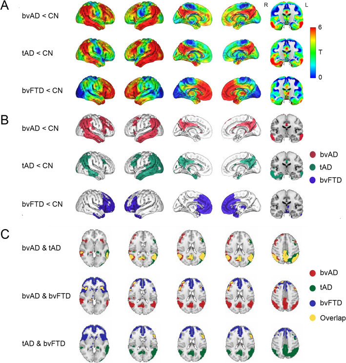
Results: bvAD and tAD showed predominant temporoparietal hypometabolism compared to controls, and did not differ in direct contrasts. However, overlaying statistical maps from contrasts between patients and controls revealed broader frontoinsular hypometabolism in bvAD than tAD, partially overlapping with bvFTD. bvAD showed greater anterior default mode network (DMN) involvement than tAD, mimicking bvFTD, and reduced connectivity of the posterior cingulate cortex with prefrontal regions. Analyses of WMH and subcortical volume showed closer resemblance of bvAD to tAD than to bvFTD, and larger amygdalar volumes in bvAD than tAD respectively. The top-3 discriminators for bvAD vs. bvFTD were FDG posterior-DMN-ratios (bvAD<bvFTD), MRI posterior-DMN-ratios (bvAD<bvFTD), MRI salience-network-ratios (bvAD>bvFTD, area under the curve [AUC] range 0.85-0.91, all p < 0.001). The top-3 for bvAD vs. tAD were amygdalar volume (bvAD>tAD), MRI anterior-DMN-ratios (bvAD<tAD), FDG anterior-DMN-ratios (bvAD<tAD, AUC range 0.71-0.84, all p < 0.05).

Conclusions: Subtle frontoinsular hypometabolism and anterior DMN involvement may underlie the prominent behavioral phenotype in bvAD.

Keywords: Alzheimer’s disease; Behavior; Frontotemporal dementia; MRI; PET.


Conflict of interest statement

The authors declare that they have no competing interests.

Figures

Fig. 1
Fig. 1
Patterns of hypometabolism of patients versus cognitively normal controls. a Surface rendering of T-maps showing hypometabolic regions in patient groups compared to cognitively healthy controls. Contrasts were adjusted for age and sex. b Surface rendering of significant voxels from contrasts between patients and controls, displayed at p < 0.05, family-wise error corrected, extent threshold k = 50. c Overlay of the T-maps from the voxel-wise comparison of FDG-PET SUVr between patients and controls. Overlays are displayed at p < 0.05, family-wise error corrected, extent threshold k = 50. Cerebellum was removed for visualization purposes
Fig. 2
Fig. 2
Connectivity patterns across groups. a The covariance maps across bvAD patients per network in red and the overlap with standard network templates by Shirer et al. [29] in yellow on the left. On the right, the goodness-of-fit score per network per patient group is depicted, showing the mean T-score of the covariance map within the network template − the mean T-score of the covariance map outside the network template. Covariance maps were obtained at puncorrected < .001, without using an extent threshold, and corrected for age and sex. b Differences in connectivity between the seed region of the default mode network (posterior cingulate cortex, top left) and the seed region of the salience network (frontoinsula, bottom left) with the rest of the brain between patient groups. Results were obtained both at puncorrected < .001 and puncorrected < .05, without using an extent threshold, and corrected for age and sex. On the right, the relationships between the SUVR in the seed region and the residualized SUVR (corrected for age and sex) in the most significant cluster resulting from the patients vs patients connectivity contrasts. pDMN posterior default mode network, aDMN anterior default mode network, SAL salience network, ECN executive control network
Fig. 3
Fig. 3
Subcortical gray matter volumes and regional white matter hyperintensity volumes across diagnostic groups. a Subcortical gray matter volumes, displayed in cm3. Error bars indicate standard deviations. *p < 0.05, **p < 0.001, Bonferroni corrected (black indicating patient contrasts, while gray represents patient vs. control contrasts). Green structures in the MRI template indicate the caudate nucleus, dark blue structures indicate the putamen, red structures indicate the globus pallidus, yellow indicate the thalamus, and light blue structures indicate the amygdala. b Regional white matter hyperintensity volumes. In this plot, the angular sections correspond to different lobes while the concentric rings represent equidistant layers of white matter. Radius increases with the distance to the ventricles (center layer: periventricular – outer layer: juxtacortical). Grayed-out regions indicate regions where the difference when compared to the control group did not reach significance. Colored regions from light yellow to red indicate the multiplicative factor when compared to control group after correction for sex, scanner field strength, and total intracranial volume
Fig. 4
Fig. 4
Top-5 discriminators for each contrast. The area-under-the-curve (AUC) and its 95% confidence interval are presented. MRI FTD = FTD signature region gray matter volume on MRI, consisting of the anterior cingulate, frontoinsula, striatum, and frontopolar regions [39, 40]; MRI AMYG = bilateral amygdala gray matter volume on MRI; MRI HIP = bilateral hippocampus gray matter volume; MRI TPC = temporoparietal gray matter volume; MRI PAR = parietal gray matter volume; MRI aDMN = gray matter volume within the anterior default mode network, divided by the gray matter volume without the anterior default mode network based on MRI; MRI pDMN = gray matter volume within the posterior default mode network, divided by the gray matter volume without the posterior default mode network based on MRI; MRI SAL = gray matter volume within the salience network, divided by the gray matter volume without the salience network based on MRI; FDG TPC = temporoparietal cortex metabolism on FDG-PET; FDG PAR = parietal cortex metabolism on FDG-PET; FDG pDMN = glucose metabolism within the posterior default mode network, divided by the glucose metabolism without the posterior default mode network on FDG-PET; FDG aDMN = glucose metabolism within the anterior default mode network, divided by the glucose metabolism without the anterior default mode network on FDG-PET; FDG SAL = glucose metabolism within the salience network, divided by the glucose metabolism without the salience network on FDG-PET; WMH BGIT = white matter hyperintensity volume in the basal ganglia and infratentorial regions

References

    1. Ossenkoppele R, Pijnenburg YA, Perry DC, Cohn-Sheehy BI, Scheltens NM, Vogel JW, et al. The behavioural/dysexecutive variant of Alzheimer’s disease: clinical, neuroimaging and pathological features. Brain. 2015;138(Pt 9):2732–2749. doi: 10.1093/brain/awv191. - DOI - PMC - PubMed
    1. Blennerhassett R, Lillo P, Halliday GM, Hodges JR, Kril JJ. Distribution of pathology in frontal variant Alzheimer’s disease. J Alzheimer's Dis. 2014;39(1):63–70. doi: 10.3233/JAD-131241. - DOI - PubMed
    1. Kawakatsu S, Kobayashi R, Hayashi H. Typical and atypical appearance of early-onset Alzheimer’s disease: a clinical, neuroimaging and neuropathological study. Neuropathol. 2017;37(2):150–173. doi: 10.1111/neup.12364. - DOI - PubMed
    1. Phillips JS, Da Re F, Dratch L, Xie SX, Irwin DJ, McMillan CT, et al. Neocortical origin and progression of gray matter atrophy in nonamnestic Alzheimer’s disease. Neurobiol Aging. 2018;63:75–87. doi: 10.1016/j.neurobiolaging.2017.11.008. - DOI - PMC - PubMed
    1. Wong S, Strudwick J, Devenney E, Hodges JR, Piguet O, Kumfor F. Frontal variant of Alzheimer’s disease masquerading as behavioural-variant frontotemporal dementia: a case study comparison. Neurocase. 2019;25(1–2):48–58. doi: 10.1080/13554794.2019.1609523. - DOI - PubMed

Publication types

MeSH terms