. 2020 Nov 14;26(1):110.
doi: 10.1186/s10020-020-00238-3.

Increased H3K4me3 methylation and decreased miR-7113-5p expression lead to enhanced Wnt/β-catenin signaling in immune cells from PTSD patients leading to inflammatory phenotype

Affiliations

Increased H3K4me3 methylation and decreased miR-7113-5p expression lead to enhanced Wnt/β-catenin signaling in immune cells from PTSD patients leading to inflammatory phenotype

Marpe Bam et al. Mol Med. .

Abstract

Background: Posttraumatic stress disorder (PTSD) is a psychiatric disorder accompanied by chronic peripheral inflammation. What triggers inflammation in PTSD is currently unclear. In the present study, we identified potential defects in signaling pathways in peripheral blood mononuclear cells (PBMCs) from individuals with PTSD.

Methods: RNAseq (5 samples each for controls and PTSD), ChIPseq (5 samples each) and miRNA array (6 samples each) were used in combination with bioinformatics tools to identify dysregulated genes in PBMCs. Real time qRT-PCR (24 samples each) and in vitro assays were employed to validate our primary findings and hypothesis.

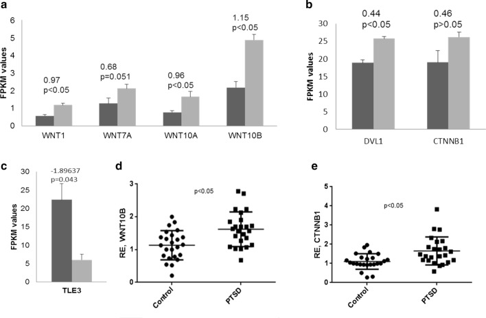
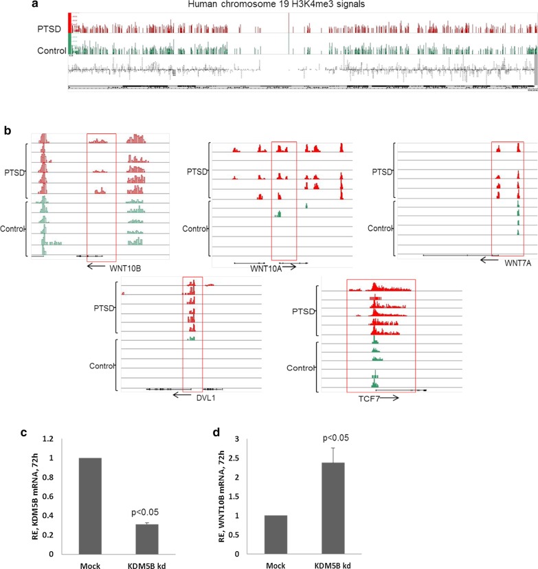
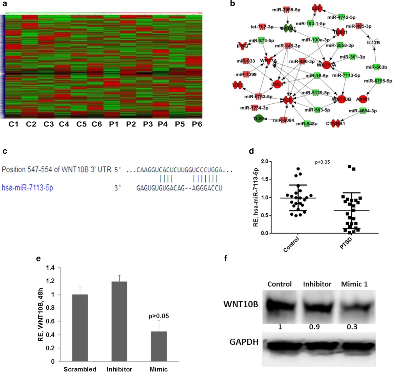
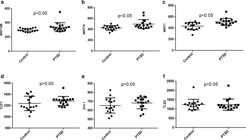
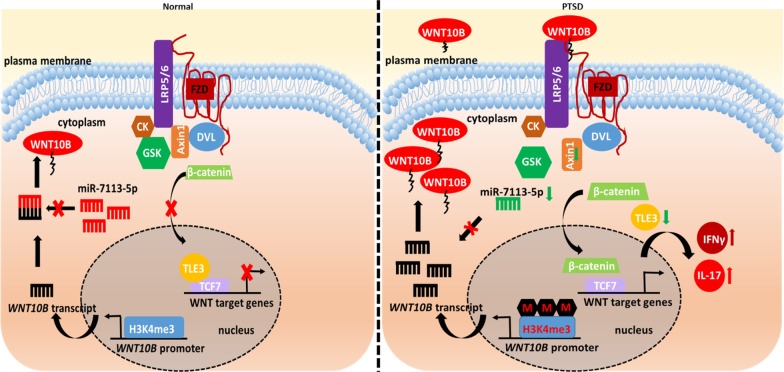
Results: By RNA-seq analysis of PBMCs, we found that Wnt signaling pathway was upregulated in PTSD when compared to normal controls. Specifically, we found increased expression of WNT10B in the PTSD group when compared to controls. Our findings were confirmed using NCBI's GEO database involving a larger sample size. Additionally, in vitro activation studies revealed that activated but not naïve PBMCs from control individuals expressed more IFNγ in the presence of recombinant WNT10B suggesting that Wnt signaling played a crucial role in exacerbating inflammation. Next, we investigated the mechanism of induction of WNT10B and found that increased expression of WNT10B may result from epigenetic modulation involving downregulation of hsa-miR-7113-5p which targeted WNT10B. Furthermore, we also observed that WNT10B overexpression was linked to higher expression of H3K4me3 histone modification around the promotor of WNT10B. Additionally, knockdown of histone demethylase specific to H3K4me3, using siRNA, led to increased expression of WNT10B providing conclusive evidence that H3K4me3 indeed controlled WNT10B expression.

Conclusions: In summary, our data demonstrate for the first time that Wnt signaling pathway is upregulated in PBMCs of PTSD patients resulting from epigenetic changes involving microRNA dysregulation and histone modifications, which in turn may promote the inflammatory phenotype in such cells.


Conflict of interest statement

The authors declare that they have no conflict of interest.

Figures

Fig. 1
Fig. 1
RNA-seq analysis using PBMCs from PTSD and control participants. Five control and 5 PTSD patient samples each were included for the analysis using total RNA from PBMCs as described previously by us (Zhou et al. 2014). The expression levels of different genes including WNT ligands (a) and WNT signaling pathway (b), shown as FPKM values. Black and grey bars represent control and PTSD samples, respectively. The values above the bars indicate log2 fold change as obtained after RNAseq data analysis. c Expression level of TLE3 in control and PTSD patients. The transcript level of WNT10B and CTNNB1was validated by qRT-PCR in 24 control and PTSD samples each (d, e). The p values were derived from the RNA-seq data analysis tool (ac) and Student’s t test (d, e)
Fig. 2
Fig. 2
ChIP-seq analysis for H3K4me3 marks. Six PTSD and 6 control samples were analyzed. In panel (a), a representative H3K4me3 ChIP-seq signal obtained after visualization of the sequencing data in Integrated Genome Browser (IGB) for human chromosome 19 is shown. b H3K4me3 signal around 5 genes of the WNT signaling pathway in 6 controls and 6 PTSD samples. One horizontal line represents a sample. c KDM5B was knocked down by siRNA in THP1 cells and confirmed by qRT-PCR after 72 h. The bar graph shows level of KDM5B transcripts in siRNA transfected cells. d WNT10B transcript level in KDM5B knocked down cells after 72 h. (RE relative expression)
Fig. 3
Fig. 3
Micro-RNA expression analysis in PTSD patients. Microarray was performed on 6 controls and 6 PTSD patients each. a Heat map showing the normalized expression intensities of the miRNAs in controls and PTSD patients (C1–C6: Control, P1–P6: PTSD). b Dysregulated miRNAs were used for miRNA target gene identification in Ingenuity Pathway Analysis tool, Qiagen. The green color miRNAs are down regulated in PTSD and red color indicate those that are upregulated. c Nucleotide pairing schema for miR-7113-5p and WNT10B 3′UTR obtained from TargetScan. d RT-qPCR validation of hsa-miR-7113-5p expression in 24 controls and PTSD patients each. WNT10B transcript (e) and protein (f) levels in THP1 cells 48 h after transfection with hsa-miR-7113-5p mimics. The values in panel f indicate the normalized ratios of the different samples
Fig. 4
Fig. 4
WNT10B-mediated effect on expression of proinflammatory genes. PBMCs from control individuals were cultured for indicated time with/without rh-WNT10B (200 ng/ml) after pre-activation of the cells with PMA (200 µM). ad qRT-PCR results showing expression of Tbx-21 and IFNγ after WNT10B addition compared to activated PBMCs after the indicated time of culture. e IFNγ was measured in the culture supernatants after 24 h of WNT10B addition. f Inflammatory gene expression levels after blocking WNT signaling by IWR1 for the indicated time period. The first vertical bars represent controls against which the experiments were compared for fold change calculation. (IWR: IWR1, WNT signaling inhibitor)
Fig. 5
Fig. 5
WNT pathway gene expression analysis from GEO Dataset. We compared the expression levels of WNT pathway genes in a GEO dataset (GSE860, Dataset 2), with data obtained from PBMCs of PTSD and controls by microarray, to confirm the observations in our dataset obtained from samples we collected. af WNT pathway genes expression levels in Dataset 2. ac corresponds to different WNT ligands upregulated in PTSD. One symbol represents an individual. The expression level corresponds to the values obtained from the analysis performed on GEO2R in the GEO dataset link. The p values were obtained by performing Student’s t test on the values obtained from the link
Fig. 6
Fig. 6
Schematic showing our working hypothesis of WNT10B elevation leading to increased inflammatory gene expression because of histone modification and miRNA expression alteration in PTSD

References

    1. Al-Harthi L. Wnt/beta-catenin and its diverse physiological cell signaling pathways in neurodegenerative and neuropsychiatric disorders. J Neuroimmune Pharmacol. 2012;7:725–730. doi: 10.1007/s11481-012-9412-x. - DOI - PMC - PubMed
    1. American Psychiatric Association . DSM-5 Task Force: diagnostic and statistical manual of mental disorders: DSM-5. Washington, D.C.: American Psychiatric Association; 2013.
    1. Bam M, Yang X, Zhou J, et al. Evidence for epigenetic regulation of pro-inflammatory cytokines, interleukin-12 and interferon gamma, in peripheral blood mononuclear cells from PTSD patients. J Neuroimmune Pharmacol. 2016;11:168–181. doi: 10.1007/s11481-015-9643-8. - DOI - PMC - PubMed
    1. Bam M, Yang X, Zumbrun EE, et al. Dysregulated immune system networks in war veterans with PTSD is an outcome of altered miRNA expression and DNA methylation. Sci Rep. 2016;6:31209. doi: 10.1038/srep31209. - DOI - PMC - PubMed
    1. Bam M, Yang X, Zumbrun EE, et al. Decreased AGO2 and DCR1 in PBMCs from War Veterans with PTSD leads to diminished miRNA resulting in elevated inflammation. Transl Psychiatry. 2017;7:e1222. doi: 10.1038/tp.2017.185. - DOI - PMC - PubMed

Publication types

MeSH terms