Review
. 2021 Jan;70(1):19-29.
doi: 10.1016/j.alit.2020.10.002. Epub 2020 Nov 12.

How to detect eosinophil ETosis (EETosis) and extracellular traps

Affiliations
Review

How to detect eosinophil ETosis (EETosis) and extracellular traps

Mineyo Fukuchi et al. Allergol Int. 2021 Jan.

Abstract

Eosinophils are short-lived and comprise only a small population of circulating leukocytes; however, they play surprisingly multifunctional roles in homeostasis and various diseases including allergy and infection. Recent research has shed light on active cytolytic eosinophil cell death that releases eosinophil extracellular traps (EETs) and total cellular contents, namely eosinophil extracellular trap cell death (EETosis). The pathological contribution of EETosis was made more cogent by recent findings that a classical pathological finding of eosinophilic inflammation, that of Charcot-Leyden crystals, is closely associated with EETosis. Currently no gold standard methods to identify EETosis exist, but "an active eosinophil lysis that releases cell-free granules and net-like chromatin structure" appears to be a common feature of EETosis. In this review, we describe several approaches that visualize EETs/EETosis in clinical samples and in vitro studies using isolated human eosinophils. EETs/EETosis can be observed using simple chemical or fluorescence staining, immunostaining, and electron microscopy, although it is noteworthy that visualization of EETs is greatly changed by sample preparation including the extracellular space of EETotic cells and shear flow. Considering the multiple aspects of biological significance, further study into EETs/EETosis is warranted to give a detailed understanding of the roles played in homeostasis and disease pathogenesis.

Keywords: Charcot-leyden crystal; EETosis; EETs; Eosinophil; NETs.


Figures

Fig. 1.
Fig. 1.
“EOSMAN” demonstrates eosinophil fates. Eosinophils normally comprise a small fraction of circulating blood leukocytes, although they can play critical roles in homeostasis and certain pathological conditions. Eosinophil biology includes production, longevity, accumulation, activation status, and secretory functions. We have created a cartoon character, EOSMAN, that might promote public awareness, research into eosinophil biology, and aid mutual understanding between physicians and patients with eosinophilic diseases.
Fig. 2.
Fig. 2.
Cytolytic EETosis releases free granules and EETs. (A) Temporal course of EETosis. Human eosinophils activated by stimuli, initially attached to the culture plate, followed by nuclear rounding and chromatin decondensation. Extracellular vesicles are also released from plasma membrane protrusions and occasionally intracellular Charcot-Leyden crystals (CLCs) are formed before plasma membrane dissolution. Finally, nuclear and plasma membranes are both disintegrated and net-like chromatin structures are liberated. EETs can be spread by shear stress. (B) Diff-quick staining of EETosis. Plasma membrane disintegration, net-like nuclear contents, and extracellular granules are observed. Eosinophils were stimulated with 1 ng/ml of IL-5 and 1 μM platelet-activating factor for 3 h in RPMI 1640 with 0.3% bovine serum albumin. (40 × objective; scale bar shows 50 μm). (C) Immunostaining for granule proteins of released EETs. Purified human eosinophils were stimulated with A23187 for 60 min to induce EETosis, then stained with anti-MBP Ab (green) for granule proteins. Propidium iodine (PI; red) was used for staining DNA. The images were obtained using fluorescence microscopy (100 × objective; scale bar shows 10 μm). Detailed methods are described in Reference.
Fig. 3.
Fig. 3.
Characteristics of EETs in vitro. (A) SYTOX staining and effect of shear stress. Eosinophils were stimulated with PMA for 3 h in the presence of cell-impermeable DNA dye SYTOX green dye (>90% of eosinophils were SYTOX positive). Under static culture conditions, net-like EETs were barely identified because of adherence to originating cells. (B) After overnight incubation (>99% of eosinophils were SYTOX positive), the culture plate was shaken using a plate shaker. Large aggregated EETs were then visualized. (Ci) Bright field image of microbeads trapped by EETs (4 × objective). After EETosis induction by stimulation with 2 μM A23187, 1 μm fluorescence beads (red, sulfate-modified beads; green, amine-modified beads) were added to the medium followed by induction of aggregation using a plate shaker. (Cii) A fluorescence image is the boxed area seen at higher magnification. Hydrophobic sulfate beads bound preferentially to EETs. (D) Ultrastructure of sulfate bead-trapping EETs. (E) EETs bound to E. coli. Detailed methods are described in Reference.
Fig. 4.
Fig. 4.
Aggregated EETs in eosinophilic exudate. Pseudocolor representation of EETs evaluated throughout the depths of eosinophilic mucin obtained from a patient with eosinophilic chronic rhinosinusitis (chronic rhinosinusitis with nasal polyps). Fixed eosinophilic mucin was stained for DNA using SYTOX green. Depth-level serial Z-stack images were pseudocolored according to the indicated color depth scale and projected to 3D using Zeiss LSM software (60 × objective). Detailed methods are described in Reference.
Fig. 5.
Fig. 5.
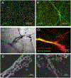
Extracellular histone staining. (A) Isolated eosinophils were stimulated with PMA for 15 min, followed by fixation (without permeabilization) and immunostained for histone H1 and DNA. Merged image of histone H1 (green), fluorescence DNA dye propidium iodine (red) and differential interference contrast (DIC) were obtained by confocal microscopy (100 × objective). Because anti-histone H1 antibodies do not penetrate intact nuclear and plasma membranes, cells were positive for DNA only. (B) After 120 min stimulation with PMA, eosinophils were similarly stained for histone H1 and DNA. Released EETs from lytic eosinophils were stained with anti-histone H1 antibodies. Note that EETs were only from lytic cells, but often attached to the neighboring intact cells (arrows). Detailed methods are described in Reference. (C) Bronchial secretion smear obtained from a patient with allergic bronchopulmonary aspergillosis was fixed and stained with histone H1 (green) and DNA (Hoechst33342, blue). EETs and chromatolytic cells were stained with anti-histone H1 antibodies. The image was obtained with a fluorescence confocal microscope using a larger pinhole diameter because of the sample thickness (20 × objective). (D) 3D reconstructed Z-stack confocal image of a bronchial secretion smear obtained from a patient with allergic bronchopulmonary aspergillosis.
Fig. 6.
Fig. 6.
Citrullinated histone staining. (A) Isolated eosinophils were stimulated with PMA for 15 min, followed by fixation (without permeabilization) and stained for citrullinated histone H3 (CitH3, green) and DNA (blue, Hoechst33342 DNA dye). The DIC image was merged (20 × objective). (B) After 120 min stimulation with PMA, eosinophils were similarly stained for citrullinated histone H3 and DNA. (Ci) Section of a bronchial mucus plug obtained from a patient with allergic bronchopulmonary aspergillosis was stained for citrullinated histone H3 (green) and DNA (blue) (4 × objective). (Cii) The boxed area in (Ci) was seen at higher magnification. (Di, Dii) A section identical to that in (Ci, Cii) was further stained with hematoxylin and eosin. Note that chromatolytic eosinophils and EETs were stained with citrullinated histone H3, but intact nuclei were not. Detailed methods are described in Reference.
Fig. 7.
Fig. 7.
Ultrastructural morphologies of EETosis. (A) Isolated blood eosinophils were observed using transmission electron microscopy. Nucleus (N) showed heterochromatin and electron-dense granules (Gr). (B) Eosinophils were stimulated with a combination of IL-5 and platelet activating factor for 3 h. Chromatolytic nucleus (N) and free extracellular granules (FEGs) were evident. (C) Nerve tissue sample obtained from a patient with eosinophilic granulomatosis with polyangiitis. EETotic eosinophils with chromatolytic nucleus (N) and FEGs are indicated. (D) Electron micrograph for lymph node obtained from a patient with hypereosinophilic syndrome. Abundant CLCs and FEGs were present.
Fig. 8.
Fig. 8.
Detection of lytic eosinophils using galectin-10 and MBP staining. (A) Isolated eosinophils were stimulated with PMA for 15 min, followed by fixation (with permeabilization) and stained for cytoplasmic galectin-10 (green), granular protein MBP (red), and DNA (blue). Merged immunofluorescence staining and DIC images were obtained by confocal microscopy. (B) After 120 min stimulation with PMA, eosinophils were similarly stained for galectin-10 and MBP. Notably, nuclear lobulation was lost in lytic galectin-10 negative cells and MBP were not co-localized with DNA. Bipyramidal Charcot-Leyden crystals were occasionally observed in EETotic cells (arrows). (C) Section of a nasal polyp obtained from a patient with eosinophilic chronic rhinosinusitis (chronic rhinosinusitis with nasal polyps) was stained for galectin-10 (green), MBP (red), and DNA (blue) (4 × objective). (D) At higher magnification (100 × objective), CLCs were associated with lytic eosinophils (arrows). (E) A section identical to that in (C) was further stained with H&E. Massive MBP deposition was observed even in eosinophils that were no longer recognizable.

References

    1. Weller PF, Spencer LA. Functions of tissue-resident eosinophils. Nat Rev Immunol 2017;17:746–60. - PMC - PubMed
    1. Nagase H, Ueki S, Fujieda S. The roles of IL-5 and anti-IL-5 treatment in eosinophilic diseases: asthma, eosinophilic granulomatosis with polyangiitis, and eosinophilic chronic rhinosinusitis. Allergol Int 2020;69:178–86. - PubMed
    1. Persson CG, Erjefalt JS. Eosinophil lysis and free granules: an in vivo paradigm for cell activation and drug development. Trends Pharmacol Sci 1997;18: 117–23. - PubMed
    1. Ueki S, Melo RC, Ghiran I, Spencer LA, Dvorak AM, Weller PF. Eosinophil extracellular DNA trap cell death mediates lytic release of free secretion-competent eosinophil granules in humans. Blood 2013;121:2074–83. - PMC - PubMed
    1. Muniz VS, Silva JC, Braga YAV, Melo RCN, Ueki S, Takeda M, et al. Eosinophils release extracellular DNA traps in response to Aspergillus fumigatus. J Allergy Clin Immunol 2018;141:571–85. e7. - PubMed