Review
. 2021 Jan;37(1):48-64.
doi: 10.1016/j.pt.2020.10.005. Epub 2020 Nov 11.

Ivermectin: An Anthelmintic, an Insecticide, and Much More

Affiliations
Review

Ivermectin: An Anthelmintic, an Insecticide, and Much More

Richard J Martin et al. Trends Parasitol. 2021 Jan.

Abstract

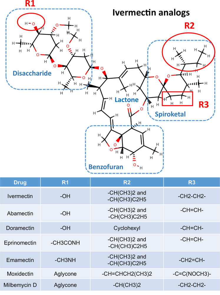
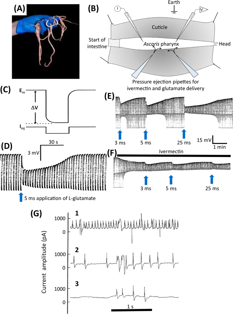
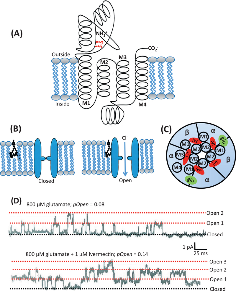
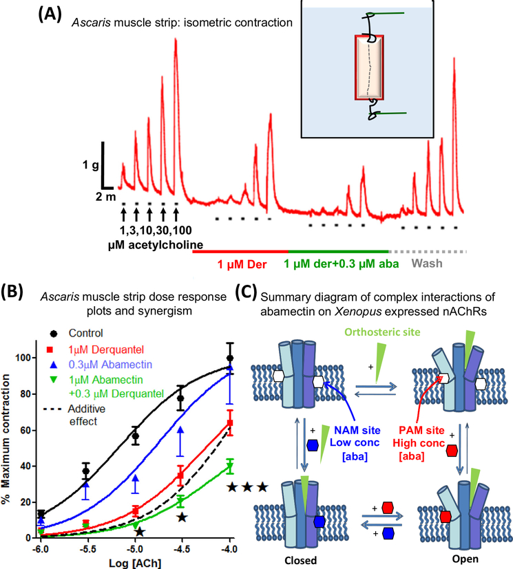
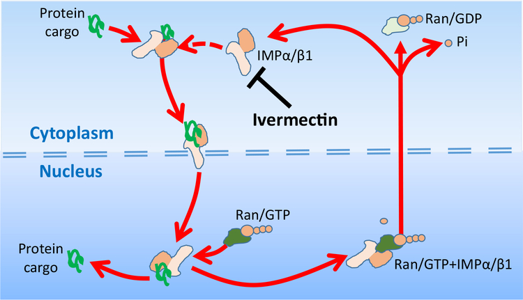
Here we tell the story of ivermectin, describing its anthelmintic and insecticidal actions and recent studies that have sought to reposition ivermectin for the treatment of other diseases that are not caused by helminth and insect parasites. The standard theory of its anthelmintic and insecticidal mode of action is that it is a selective positive allosteric modulator of glutamate-gated chloride channels found in nematodes and insects. At higher concentrations, ivermectin also acts as an allosteric modulator of ion channels found in host central nervous systems. In addition, in tissue culture, at concentrations higher than anthelmintic concentrations, ivermectin shows antiviral, antimalarial, antimetabolic, and anticancer effects. Caution is required before extrapolating from these preliminary repositioning experiments to clinical use, particularly for Covid-19 treatment, because of the high concentrations of ivermectin used in tissue-culture experiments.

Keywords: anthelmintic; anticancer; antimalaria; antivirus; insecticide; ivermectin.


Figures

Figure 1.
Figure 1.. Structure of Ivermectin and Analogs.
The molecule is divided into four regions: the disaccharide, the lactone, the spiroketal, and the benzofuran. Different modifications to this structure at the positions R1, R2, and R3 give rise to ivermectin, abamectin, doramectin, eprinomectin, emamectin, moxidectin, and milbemycin.
Figure 2.
Figure 2.. Electrophysiologic Effects of Ivermectin in Parasitic Nematodes.
(A) Ascaris suum is a large nematode that is tractable for electrophysiologic recordings. (B) A diagram of the dissected A. suum pharynx. Two glass micropipettes are placed in the pharynx, one for injecting rectangular current pulses (I) and one for recording the membrane potential (V). Two ‘puffing’ microelectrodes are used to apply, separately, glutamate or ivermectin at different time intervals. (C) Higher time resolution recording showing the injection of a hyperpolarizing rectangular current pulse (Iinj) and the membrane potential (Em) and response to the injected current (ΔV) allowing the input conductance (= 1/resistance) of the pharynx to be measured. (D) A 5 ms puff of 0.5 M glutamate produces a hyperpolarization due to the entry of Cl (upper envelope of the trace) and a reduction in the width of the trace as the size of the membrane potential change (ΔV) is reduced as the glutamate-gated chloride (GluCl) channels open and increase the conductance of the pharyngeal muscle membrane. (E) Effects of glutamate. The trace shows the membrane potential and input conductance responses to 3 ms, 5 ms, and 25 ms ‘puffs’ of 0.5 M glutamate from a micropipette, which produces maximal changes in input conductance of 16 μS, 33 μS, and 60 μS, respectively. Longer applications of glutamate did not produce an increase in response in this experiment, showing that the 25 μs response led to a maximum response. F) The effect of a continuous ‘puff’ of 100 μM ivermectin applied also by pressure ejection from a micropipette. Once the conductance change produced by the ivermectin reached a stable position of 32.7 μS, glutamate was again applied in the same controlled way using the same application times; this produced a maximum conductance change of 28 μS with a 25 ms puff application. The maximum conductance change obtained with the coapplication of glutamate and ivermectin was 60 μS, virtually the same conductance (cf. 60 μS) change produced after a high-dose application of glutamate. The fact that coapplication did not produce an effect greater than the high dose of glutamate was interpreted as indicating that these two substances activate the same ion channel – because application of two separate ion channels by glutamate and ivermectin at high concentrations would produce an additive response. (G) Electropharyngeograms from Trichostrongylus colubriformis A before, 5 min after, and 15 min after adding 100 nM ivermectin to the bath (from [106]). A patch-pipette was used to record the currents elicited by the pumping pharynx by sucking the head of the worm into the patch-pipette. (1) Control recording before application of 100 nM ivermectin; (2) 5 min after application of 100 nM ivermectin; (3) 15 min after application of 100 nM ivermectin.
Figure 3.
Figure 3.. Glutamate-gated Chloride (GluCl) Channel Structure, Ivermectin and Glutamate Binding Sites, and Channel Currents.
(A) Diagram of a GluCl channel subunit. It is about 500 amino acids long and begins at the N terminal on the extracellular face of the membrane. The subunits that contain vicinal cysteines are recognized as α-subunits. Each subunit has four α-helical transmembrane regions: M1, M2, M3, and M4. The M2 region lines the pore of the transmembrane ion channel. Between the M3 and M4 regions there is an extended cytoplasmic loop. The C terminal is also found at the extracellular surface of the membrane. (B) Five subunits are arranged like the staves of a barrel around the central pore of the ion channel to form a pentomer. Ivermectin binds to the channel in the transmembrane region of the ion channel in the outer bilayer of the lipid bilayer membrane and can open the channel. (C) The five subunits of the GluCl channel viewed from above. If the five subunits of the ion channel are all the same, then the ion channel receptor is referred to as homomeric. If it is made of nonidentical subunits it is referred to as heteromeric. The orthosteric ligand-binding site, where glutamate binds, is located between two adjacent subunits (green) and in the extracellular region of the ion channel. The ivermectin binding site is deep in the ion channel and is involved in contact with the M2 region near the pore, and it contacts the M3 region as well. (D) The upper trace shows a cell-attached patch recording at −50 mV with 800 μM glutamate in the patch-pipette. The rectangular current steps representing the opening and closing of GluCl channels can be seen. Each current step is 2 pA (10−12 A). The probability of the channels being open (pOpen) is 0.08, that is, each channel was open for 8% of the time. The lower trace shows the effect of adding 1 μM ivermectin to the bath, and followed after a few minutes, allowing time for ivermectin to move into and along the membrane, the number of GluCl opening increased as did the probability of the channel being open. It increased to 0.14, or 14% of the time. The opening of many ion channels together allows more Cl current to flow through the membranes and to increase the conductance of the membrane.
Figure 4.
Figure 4.. Interactions of Abamectin and Derquantel on Ascaris Muscle Strip Contraction and Summary Diagram of Complex Effects of Abamectin on Nicotinic Acetylcholine Receptors (nAChRs).
(A) Isometric contraction of Ascaris suum muscle strips produced by application of increasing concentrations of acetylcholine and antagonism by 1 μM derquantel (red bar), 1 μM derquantel + 0.3 μM abamectin (green bar), and wash (gray bar). Note that derquantel decreases the responses to acetylcholine and that the addition of abamectin increases the inhibition. (B) An A. suum muscle strip concentration–contraction–response plot of acetylcholine showing mean ± S.E. bars (n = 11). Control (black); in the presence of 1 μM derquantel (red); 1 μM derquantel + 0.3 μM abamectin (green); and wash (blue). Note that abamectin increases the inhibition produced by derquantel. The inhibitory effect of abamectin and derquantel is greater than an additive effect, dotted line [37]. (C) Model of ligand sites of action showing complex effects on heteromeric ion channels. The cholinergic anthelmintic agonists bind to the orthosteric sites, opening the channel. Low concentrations of abamectin (0.03 and 0.1 μM) bind to a negative allosteric site (NAM) in the lipid phase of the channel, inhibiting opening. Higher concentrations of abamectin (0.3, 1, and 10 μM) bind to a positive allosteric site (PAM), increasing opening [35].
Figure 5.
Figure 5.. Ivermectin Inhibits Nuclear Import of Selected Proteins, Including Some Virus Proteins.
Illustrations of how ivermectin binds to the heterodimer protein importin, IMPα/β1, to inhibit the binding of cargo proteins that are carried through the nuclear pore by IMPα/β1 into the nucleus. The cargo protein is recognized by the IMPα/β1 protein by the α/β by a nuclear localization signal (NLS), short sequences of basic amino acids either alone or separated by a linker region of 10–12 amino acids. The import process is energy-dependent, involving GTP and its hydrolysis. Once through the nuclear pore, the protein is released when RanGTP binds to IMPα/β1 to allow release of the cargo into the nucleus. The empty IMPα/β1 is then shuttled back out through the nuclear pore to continue the cycle. GDP,; GTP,; Ran,.

References

    1. Canga AG et al. (2008) The pharmacokinetics and interactions of ivermectin in humans – A mini-review. AAPS J. 10, 42–46 - PMC - PubMed
    1. González Canga A et al. (2009) The pharmacokinetics and metabolism of ivermectin in domestic animal species. Vet. J. 179, 25–37 - PubMed
    1. Merck (2019) Stomectol safety data sheet. p. 7
    1. Bauck S (1987) Ivermectin toxicity in small animals. Can. Vet. J. 28, 563–564 - PMC - PubMed
    1. Campbell WC et al., eds (1989) Ivermectin and Abamectin, Springer-Verlag

Publication types